Fibronectin Rabbit mAb[Y82N]Cat NO.: A51445
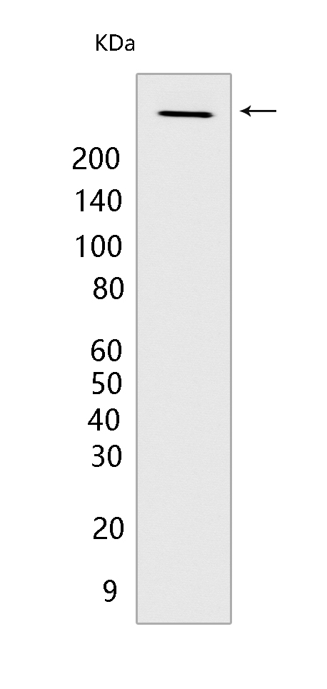
Western blot(SDS PAGE) analysis of extracts from HepG2 cells.Using Fibronectin Rabbit mAb IgG [Y82N] at dilution of 1:1000 incubated at 4℃ over night.
Product information
Protein names :FN1,FN,FINC_HUMAN,Fibronectin [Cleaved into: Anastellin,Ugl-Y1,Ugl-Y2,Ugl-Y3]
UniProtID :P02751
MASS(da) :272,320
MW(kDa) :263kDa
Form :Liquid
Purification :Protein A purification
Host :Rabbit
Isotype :IgG
sensitivity :Endogenous
Reactivity :Human,Mouse
- ApplicationDilution
- 免疫印迹(WB)1:1000-2000,
- 免疫荧光(ICC/IF) 1:100
- The optimal dilutions should be determined by the end user
Specificity :Antibody is produced by immunizing animals with a synthetic peptide of human Fibronectin.
Storage :Antibody store in 10 mM PBS, 0.5mg/ml BSA, 50% glycerol. Shipped at 4°C. Store at-20°C or -80°C. Products are valid for one natural year of receipt.Avoid repeated freeze / thaw cycles.
WB Positive detected :HepG2 cells
Function : Fibronectins bind cell surfaces and various compounds including collagen, fibrin, heparin, DNA, and actin (PubMed:3024962, PubMed:3900070, PubMed:3593230, PubMed:7989369). Fibronectins are involved in cell adhesion, cell motility, opsonization, wound healing, and maintenance of cell shape (PubMed:3024962, PubMed:3900070, PubMed:3593230, PubMed:7989369). Involved in osteoblast compaction through the fibronectin fibrillogenesis cell-mediated matrix assembly process, essential for osteoblast mineralization (By similarity). Participates in the regulation of type I collagen deposition by osteoblasts (By similarity). Acts as a ligand for the LILRB4 receptor, inhibiting FCGR1A/CD64-mediated monocyte activation (PubMed:34089617).., [Anastellin]: Binds fibronectin and induces fibril formation. This fibronectin polymer, named superfibronectin, exhibits enhanced adhesive properties. Both anastellin and superfibronectin inhibit tumor growth, angiogenesis and metastasis. Anastellin activates p38 MAPK and inhibits lysophospholipid signaling..
Tissue specificity :Expressed in the inner limiting membrane and around blood vessels in the retina (at protein level) (PubMed:29777959). Plasma FN (soluble dimeric form) is secreted by hepatocytes. Cellular FN (dimeric or cross-linked multimeric forms), made by fibroblasts, epithelial and other cell types, is deposited as fibrils in the extracellular matrix. Ugl-Y1, Ugl-Y2 and Ugl-Y3 are found in urine (PubMed:17614963)..
Subcellular locationi :Secreted, extracellular space, extracellular matrix.
IMPORTANT: For western blots, incubate membrane with diluted primary antibody in 1% w/v BSA, 1X TBST at 4°C overnight.


