GluR2+GluR3 Rabbit mAb[K0Q4]Cat NO.: A68358
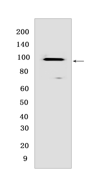
Western blot(SDS PAGE) analysis of extracts from Mouse brain tissue lysate.Using GluR2+GluR3 Rabbit mAb IgG [K0Q4] at dilution of 1:1000 incubated at 4℃ over night.
Product information
Protein names :GRIA2,GLUR2,GRIA2_HUMAN,Glutamate receptor 2
UniProtID :P42262/P42263
MASS(da) :101,157
MW(kDa) :98kDa
Form :Liquid
Purification :Protein A purification
Host :Rabbit
Isotype :IgG
sensitivity :Endogenous
Reactivity :Mouse,Rat
- ApplicationDilution
- 免疫印迹(WB)1:1000-2000
- The optimal dilutions should be determined by the end user
Specificity :Antibody is produced by immunizing animals with a synthetic peptide of Mouse GluR2+GluR3.
Storage :Antibody store in 10 mM PBS, 0.5mg/ml BSA, 50% glycerol. Shipped at 4°C. Store at-20°C or -80°C. Products are valid for one natural year of receipt.Avoid repeated freeze / thaw cycles.
WB Positive detected :Mouse brain tissue lysate
Function : Receptor for glutamate that functions as ligand-gated ion channel in the central nervous system (PubMed:31300657). It plays an important role in excitatory synaptic transmission. L-glutamate acts as an excitatory neurotransmitter at many synapses in the central nervous system. Binding of the excitatory neurotransmitter L-glutamate induces a conformation change, leading to the opening of the cation channel, and thereby converts the chemical signal to an electrical impulse. The receptor then desensitizes rapidly and enters a transient inactive state, characterized by the presence of bound agonist. In the presence of CACNG4 or CACNG7 or CACNG8, shows resensitization which is characterized by a delayed accumulation of current flux upon continued application of glutamate. Through complex formation with NSG1, GRIP1 and STX12 controls the intracellular fate of AMPAR and the endosomal sorting of the GRIA2 subunit toward recycling and membrane targeting (By similarity)..
Subcellular locationi :Cell membrane,Multi-pass membrane protein. Endoplasmic reticulum membrane,Multi-pass membrane protein. Cell junction, synapse, postsynaptic cell membrane,Multi-pass membrane protein. Cell junction, synapse, postsynaptic density membrane,Multi-pass membrane protein.
IMPORTANT: For western blots, incubate membrane with diluted primary antibody in 1% w/v BSA, 1X TBST at 4°C overnight.


