Lipocalin-2/NGAL Rabbit mAb[04TY]Cat NO.: A20336
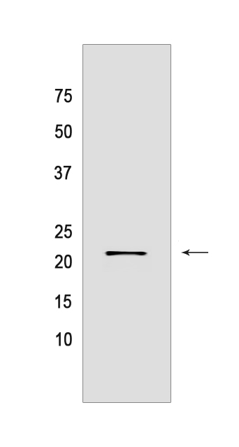
Western blot(SDS PAGE) analysis of extracts from HT-29 cells.Using Lipocalin-2/NGAL Rabbit mAb IgG [04TY] at dilution of 1:1000 incubated at 4℃ over night.
Product information
Protein names :LCN2,HNL,NGAL,NGAL_HUMAN,Neutrophil gelatinase-associated lipocalin
UniProtID :P80188
MASS(da) :22,588
MW(kDa) :22KDa
Form :Liquid
Purification :Protein A purification
Host :Rabbit
Isotype :IgG
sensitivity :Endogenous
Reactivity :Human
- ApplicationDilution
- 免疫印迹(WB)1:1000-2000
- The optimal dilutions should be determined by the end user
Specificity :Antibody is produced by immunizing animals with a synthetic peptide of human Lipocalin-2/NGAL.
Storage :Antibody store in 10 mM PBS, 0.5mg/ml BSA, 50% glycerol. Shipped at 4°C. Store at-20°C or -80°C. Products are valid for one natural year of receipt.Avoid repeated freeze / thaw cycles.
WB Positive detected :HT-29 cells
Function : Iron-trafficking protein involved in multiple processes such as apoptosis, innate immunity and renal development (PubMed:12453413, PubMed:27780864, PubMed:20581821). Binds iron through association with 2,3-dihydroxybenzoic acid (2,3-DHBA), a siderophore that shares structural similarities with bacterial enterobactin, and delivers or removes iron from the cell, depending on the context. Iron-bound form (holo-24p3) is internalized following binding to the SLC22A17 (24p3R) receptor, leading to release of iron and subsequent increase of intracellular iron concentration. In contrast, association of the iron-free form (apo-24p3) with the SLC22A17 (24p3R) receptor is followed by association with an intracellular siderophore, iron chelation and iron transfer to the extracellular medium, thereby reducing intracellular iron concentration. Involved in apoptosis due to interleukin-3 (IL3) deprivation: iron-loaded form increases intracellular iron concentration without promoting apoptosis, while iron-free form decreases intracellular iron levels, inducing expression of the proapoptotic protein BCL2L11/BIM, resulting in apoptosis (By similarity). Involved in innate immunity,limits bacterial proliferation by sequestering iron bound to microbial siderophores, such as enterobactin (PubMed:27780864). Can also bind siderophores from M.tuberculosis (PubMed:15642259, PubMed:21978368)..
Tissue specificity :Detected in neutrophils (at protein level) (PubMed:7683678, PubMed:8298140). Expressed in bone marrow and in tissues that are prone to exposure to microorganism. High expression is found in bone marrow as well as in uterus, prostate, salivary gland, stomach, appendix, colon, trachea and lung. Not found in the small intestine or peripheral blood leukocytes..
Subcellular locationi :Secreted. Cytoplasmic granule lumen. Cytoplasmic vesicle lumen.
IMPORTANT: For western blots, incubate membrane with diluted primary antibody in 1% w/v BSA, 1X TBST at 4°C overnight.


