NDRG1 Rabbit mAb[9WWP]Cat NO.: A41711
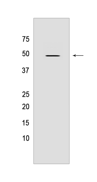
Western blot(SDS PAGE) analysis of extracts from 293T cells.Using NDRG1 Rabbit mAb IgG [9WWP] at dilution of 1:1000 incubated at 4℃ over night.
Product information
Protein names :NDRG1,CAP43,DRG1,RTP,NDRG1_HUMAN,Protein NDRG1
UniProtID :Q92597
MASS(da) :42,835
MW(kDa) :48KDa
Form :Liquid
Purification :Protein A purification
Host :Rabbit
Isotype :IgG
sensitivity :Endogenous
Reactivity :Human,Mouse,Rat
- ApplicationDilution
- 免疫印迹(WB)1:1000-2000,
- 免疫组化(IHC)1:100
- The optimal dilutions should be determined by the end user
Specificity :Antibody is produced by immunizing animals with a synthetic peptide of human NDRG1.
Storage :Antibody store in 10 mM PBS, 0.5mg/ml BSA, 50% glycerol. Shipped at 4°C. Store at-20°C or -80°C. Products are valid for one natural year of receipt.Avoid repeated freeze / thaw cycles.
WB Positive detected :293T cells
Function : Stress-responsive protein involved in hormone responses, cell growth, and differentiation. Acts as a tumor suppressor in many cell types. Necessary but not sufficient for p53/TP53-mediated caspase activation and apoptosis. Has a role in cell trafficking, notably of the Schwann cell, and is necessary for the maintenance and development of the peripheral nerve myelin sheath. Required for vesicular recycling of CDH1 and TF. May also function in lipid trafficking. Protects cells from spindle disruption damage. Functions in p53/TP53-dependent mitotic spindle checkpoint. Regulates microtubule dynamics and maintains euploidy..
Tissue specificity :Ubiquitous,expressed most prominently in placental membranes and prostate, kidney, small intestine, and ovary tissues. Also expressed in heart, brain, skeletal muscle, lung, liver and pancreas. Low levels in peripheral blood leukocytes and in tissues of the immune system. Expressed mainly in epithelial cells. Also found in Schwann cells of peripheral neurons. Reduced expression in adenocarcinomas compared to normal tissues. In colon, prostate and placental membranes, the cells that border the lumen show the highest expression..
Subcellular locationi :Cytoplasm, cytosol. Cytoplasm, cytoskeleton, microtubule organizing center, centrosome. Nucleus. Cell membrane.
IMPORTANT: For western blots, incubate membrane with diluted primary antibody in 1% w/v BSA, 1X TBST at 4°C overnight.


