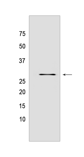
PEF1 Rabbit mAb[A8E6]Cat NO.: A16860
Western blot(SDS PAGE) analysis of extracts from Jurkat cells.Using PEF1 Rabbit mAb IgG [A8E6] at dilution of 1:1000 incubated at 4℃ over night.
Product information
Protein names :PEF1,ABP32,UNQ1845/PRO3573,PEF1_HUMAN,Peflin
UniProtID :Q9UBV8
MASS(da) :30,381
MW(kDa) :30KDa
Form :Liquid
Purification :Protein A purification
Host :Rabbit
Isotype :IgG
sensitivity :Endogenous
Reactivity :Human,Mouse,Rat
- ApplicationDilution
- 免疫印迹(WB)1:1000-2000
- The optimal dilutions should be determined by the end user
Specificity :Antibody is produced by immunizing animals with a synthetic peptide of human PEF1.
Storage :Antibody store in 10 mM PBS, 0.5mg/ml BSA, 50% glycerol. Shipped at 4°C. Store at-20°C or -80°C. Products are valid for one natural year of receipt.Avoid repeated freeze / thaw cycles.
WB Positive detected :Jurkat cells
Function : Calcium-binding protein that acts as an adapter that bridges unrelated proteins or stabilizes weak protein-protein complexes in response to calcium. Together with PDCD6, acts as calcium-dependent adapter for the BCR(KLHL12) complex, a complex involved in endoplasmic reticulum (ER)-Golgi transport by regulating the size of COPII coats (PubMed:27716508). In response to cytosolic calcium increase, the heterodimer formed with PDCD6 interacts with, and bridges together the BCR(KLHL12) complex and SEC31 (SEC31A or SEC31B), promoting monoubiquitination of SEC31 and subsequent collagen export, which is required for neural crest specification (PubMed:27716508). Its role in the heterodimer formed with PDCD6 is however unclear: some evidence shows that PEF1 and PDCD6 work together and promote association between PDCD6 and SEC31 in presence of calcium (PubMed:27716508). Other reports show that PEF1 dissociates from PDCD6 in presence of calcium, and may act as a negative regulator of PDCD6 (PubMed:11278427). Also acts as a negative regulator of ER-Golgi transport,possibly by inhibiting interaction between PDCD6 and SEC31 (By similarity)..
Subcellular locationi :Cytoplasm. Endoplasmic reticulum. Membrane,Peripheral membrane protein. Cytoplasmic vesicle, COPII-coated vesicle membrane,Peripheral membrane protein.
IMPORTANT: For western blots, incubate membrane with diluted primary antibody in 1% w/v BSA, 1X TBST at 4°C overnight.


