
AIFM2 Mouse mAb[GPAV]Cat NO.: A39646
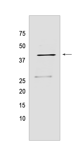
Western blot(SDS PAGE) analysis of extracts from HepG2 cells.Using AIFM2 Mouse mAb IgG [GPAV] at dilution of 1:1000 incubated at 4℃ over night.
Product information
Protein names :AIFM2,AMID,PRG3,FSP1_HUMAN,Ferroptosis suppressor protein 1
UniProtID :Q9BRQ8
MASS(da) :40,527
MW(kDa) :41kDa
Form :Liquid
Purification :Protein A purification
Host :Mouse
Isotype :IgG
sensitivity :Endogenous
Reactivity :Human
- ApplicationDilution
- 免疫印迹(WB)1:1000-2000
- The optimal dilutions should be determined by the end user
Specificity :Antibody is produced by immunizing animals with a synthetic peptide of human AIFM2.
Storage :Antibody store in 10 mM PBS, 0.5mg/ml BSA, 50% glycerol. Shipped at 4°C. Store at-20°C or -80°C. Products are valid for one natural year of receipt.Avoid repeated freeze / thaw cycles.
WB Positive detected :HepG2 cells
Function : A NAD(P)H-dependent oxidoreductase involved in cellular oxidative stress response. At the plasma membrane, catalyzes reduction of coenzyme Q/ubiquinone-10 to ubiquinol-10, a lipophilic radical-trapping antioxidant that prevents lipid oxidative damage and consequently ferroptosis. Cooperates with GPX4 to suppress phospholipid peroxidation and ferroptosis. This anti-ferroptotic function is independent of cellular glutathione levels (PubMed:31634899, PubMed:31634900). May play a role in mitochondrial stress signaling. Upon oxidative stress, associates with the lipid peroxidation end product 4-hydroxy-2-nonenal (HNE) forming a lipid adduct devoid of oxidoreductase activity, which then translocates from mitochondria into the nucleus triggering DNA damage and cell death (PubMed:26689472). Capable of DNA binding in a non-sequence specific way (PubMed:15958387)..
Tissue specificity :Detected in most normal tissues as two transcripts of 1.8 and 4.0 kb in length, respectively. Highly expressed in heart, moderately in liver and skeletal muscles, and expressed at low levels in placenta, lung, kidney, and pancreas. Both transcripts expressed following p53/TP53 induction. The shorter 1.8 kb transcript seems to be the major transcript in EB1 colon cancer cells..
Subcellular locationi :Lipid droplet. Cell membrane,Lipid-anchor. Cytoplasm. Mitochondrion membrane. Nucleus.
IMPORTANT: For western blots, incubate membrane with diluted primary antibody in 1% w/v BSA, 1X TBST at 4°C overnight.
-
About
-
Product
-
Law
-
Literature
-

WeChat scan code follow us