
ALPK1 Mouse mAb[5NJJ]Cat NO.: A24614
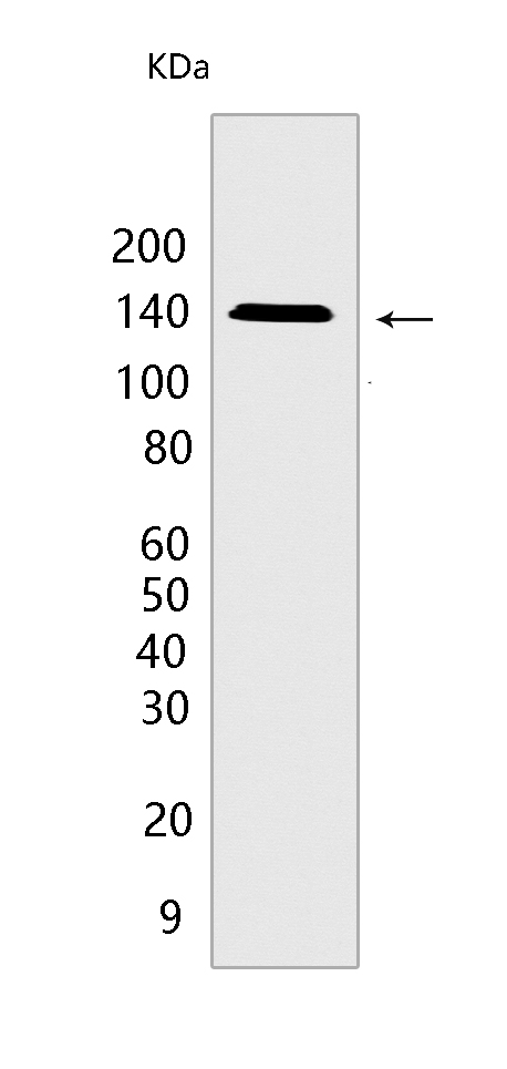
Western blot(SDS PAGE) analysis of extracts from COLO 320 cells.Using ALPK1 Mouse mAb IgG [5NJJ] at dilution of 1:1000 incubated at 4℃ over night.
Product information
Protein names :ALPK1,KIAA1527,LAK,ALPK1_HUMAN,Alpha-protein kinase 1
UniProtID :Q96QP1
MASS(da) :138,861
MW(kDa) :139kDa
Form :Liquid
Purification :Protein A purification
Host :Mouse
Isotype :IgG
sensitivity :Endogenous
Reactivity :Human,Mouse
- ApplicationDilution
- 免疫印迹(WB)1:1000-2000
- 免疫组化(IHC)1:100
- The optimal dilutions should be determined by the end user
Specificity :Antibody is produced by immunizing animals with a synthetic peptide of human ALPK1.
Storage :Antibody store in 10 mM PBS, 0.5mg/ml BSA, 50% glycerol. Shipped at 4°C. Store at-20°C or -80°C. Products are valid for one natural year of receipt.Avoid repeated freeze / thaw cycles.
WB Positive detected :COLO 320 cells
Function : Serine/threonine-protein kinase that detects bacterial pathogen-associated molecular pattern metabolites (PAMPs) and initiates an innate immune response, a critical step for pathogen elimination and engagement of adaptive immunity (PubMed:28877472, PubMed:28222186, PubMed:30111836). Specifically recognizes and binds ADP-D-glycero-beta-D-manno-heptose (ADP-Heptose), a potent PAMP present in all Gram-negative and some Gram-positive bacteria (PubMed:30111836). ADP-Heptose-binding stimulates its kinase activity to phosphorylate and activate TIFA, triggering pro-inflammatory NF-kappa-B signaling (PubMed:30111836). May be involved in monosodium urate monohydrate (MSU)-induced inflammation by mediating phosphorylation of unconventional myosin MYO9A (PubMed:27169898). May also play a role in apical protein transport by mediating phosphorylation of unconventional myosin MYO1A (PubMed:15883161). May play a role in ciliogenesis (PubMed:30967659)..
Tissue specificity :Highly expressed in liver. Expressed in the optic nerve and retinal pigmented epithelium. Lower expression is observed in the macula and extramacular retina (PubMed:30967659)..
Subcellular locationi :Cytoplasm, cytosol. Cytoplasm, cytoskeleton, spindle pole. Cytoplasm, cytoskeleton, microtubule organizing center, centrosome. Cell projection, cilium.
IMPORTANT: For western blots, incubate membrane with diluted primary antibody in 1% w/v BSA, 1X TBST at 4°C overnight.
-
About
-
Product
-
Law
-
Literature
-

WeChat scan code follow us