CD133 Mouse mAb[2W3N]Cat NO.: A10571
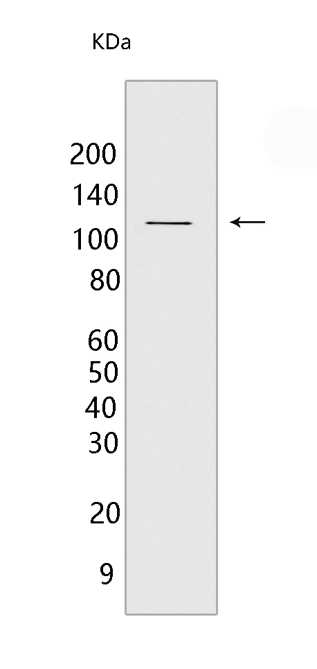
Western blot(SDS PAGE) analysis of extracts from HT-29 cells.Using CD133 Mouse mAb IgG [2W3N] at dilution of 1:1000 incubated at 4℃ over night.
Product information
Protein names :PROM1,PROML1,MSTP061,PROM1_HUMAN,Prominin-1
UniProtID :O43490
MASS(da) :97,202
MW(kDa) :115kDa,80-90kDa
Form :Liquid
Purification :Protein A purification
Host :Mouse
Isotype :IgG
sensitivity :Endogenous
Reactivity :Human
- ApplicationDilution
- 免疫印迹(WB)1:1000-2000
- 免疫组化(IHC)1:100
- The optimal dilutions should be determined by the end user
Specificity :Antibody is produced by immunizing animals with a synthetic peptide of human CD133.
Storage :Antibody store in 10 mM PBS, 0.5mg/ml BSA, 50% glycerol. Shipped at 4°C. Store at-20°C or -80°C. Products are valid for one natural year of receipt.Avoid repeated freeze / thaw cycles.
WB Positive detected :HT-29 cells
Function : May play a role in cell differentiation, proliferation and apoptosis (PubMed:24556617). Binds cholesterol in cholesterol-containing plasma membrane microdomains and may play a role in the organization of the apical plasma membrane in epithelial cells. During early retinal development acts as a key regulator of disk morphogenesis. Involved in regulation of MAPK and Akt signaling pathways. In neuroblastoma cells suppresses cell differentiation such as neurite outgrowth in a RET-dependent manner (PubMed:20818439)..
Tissue specificity :Isoform 1 is selectively expressed on CD34 hematopoietic stem and progenitor cells in adult and fetal bone marrow, fetal liver, cord blood and adult peripheral blood. Isoform 1 is not detected on other blood cells. Isoform 1 is also expressed in a number of non-lymphoid tissues including retina, pancreas, placenta, kidney, liver, lung, brain and heart. Found in saliva within small membrane particles. Isoform 2 is predominantly expressed in fetal liver, skeletal muscle, kidney, and heart as well as adult pancreas, kidney, liver, lung, and placenta. Isoform 2 is highly expressed in fetal liver, low in bone marrow, and barely detectable in peripheral blood. Isoform 2 is expressed on hematopoietic stem cells and in epidermal basal cells (at protein level). Expressed in adult retina by rod and cone photoreceptor cells (at protein level)..
Subcellular locationi :Apical cell membrane,Multi-pass membrane protein. Cell projection, microvillus membrane,Multi-pass membrane protein. Cell projection, cilium, photoreceptor outer segment. Endoplasmic reticulum. Endoplasmic reticulum-Golgi intermediate compartment.
IMPORTANT: For western blots, incubate membrane with diluted primary antibody in 1% w/v BSA, 1X TBST at 4°C overnight.


