
CD63 Mouse mAb[4D68]Cat NO.: A62713
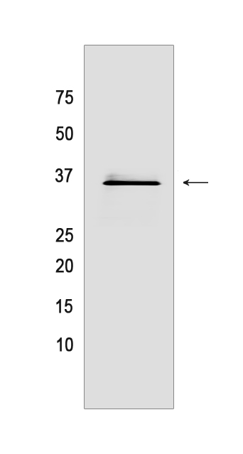
Western blot(SDS PAGE) analysis of extracts from HeLa cells.Using CD63 Mouse mAb IgG [4D68] at dilution of 1:1000 incubated at 4℃ over night.
Product information
Protein names :CD63,MLA1,TSPAN30,CD63_HUMAN,CD63 antigen
UniProtID :P08962
MASS(da) :25,637
MW(kDa) :35kDa
Form :Liquid
Purification :Protein A purification
Host :Mouse
Isotype :IgG
sensitivity :Endogenous
Reactivity :Human
- ApplicationDilution
- 免疫印迹(WB)1:1000-2000
- 免疫组化(IHC)1:100
- 免疫荧光(ICC/IF) 1:100,
- The optimal dilutions should be determined by the end user
Specificity :Antibody is produced by immunizing animals with a synthetic peptide of human CD63.
Storage :Antibody store in 10 mM PBS, 0.5mg/ml BSA, 50% glycerol. Shipped at 4°C. Store at-20°C or -80°C. Products are valid for one natural year of receipt.Avoid repeated freeze / thaw cycles.
WB Positive detected :HeLa cells
Function : Functions as cell surface receptor for TIMP1 and plays a role in the activation of cellular signaling cascades. Plays a role in the activation of ITGB1 and integrin signaling, leading to the activation of AKT, FAK/PTK2 and MAP kinases. Promotes cell survival, reorganization of the actin cytoskeleton, cell adhesion, spreading and migration, via its role in the activation of AKT and FAK/PTK2. Plays a role in VEGFA signaling via its role in regulating the internalization of KDR/VEGFR2. Plays a role in intracellular vesicular transport processes, and is required for normal trafficking of the PMEL luminal domain that is essential for the development and maturation of melanocytes. Plays a role in the adhesion of leukocytes onto endothelial cells via its role in the regulation of SELP trafficking. May play a role in mast cell degranulation in response to Ms4a2/FceRI stimulation, but not in mast cell degranulation in response to other stimuli..
Tissue specificity :Detected in platelets (at protein level). Dysplastic nevi, radial growth phase primary melanomas, hematopoietic cells, tissue macrophages..
Subcellular locationi :Cell membrane,Multi-pass membrane protein. Lysosome membrane,Multi-pass membrane protein. Late endosome membrane,Multi-pass membrane protein. Endosome, multivesicular body. Melanosome. Secreted, extracellular exosome. Cell surface.
IMPORTANT: For western blots, incubate membrane with diluted primary antibody in 1% w/v BSA, 1X TBST at 4°C overnight.
-
About
-
Product
-
Law
-
Literature
-

WeChat scan code follow us