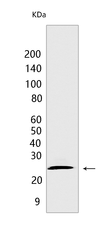
ARC Rabbit mAb [A44P]Cat NO.: A59185
Western blot(SDS PAGE) analysis of extracts from 293T cells.Using ARC Rabbit mAb [A44P] at dilution of 1:1000 incubated at 4℃ over night.
Product information
Protein names :NOL3,ARC,NOP,NOL3_HUMAN,Nucleolar protein 3
UniProtID :O60936
MASS(da) :22,629
MW(kDa) :27 kDa
Form :Liquid
Purification :Protein A purification
Host :Rabbit
Isotype :IgG
sensitivity :Endogenous
Reactivity :Human
- ApplicationDilution
- 免疫印迹(WB)1:1000-2000
- The optimal dilutions should be determined by the end user
Specificity :Antibody is produced by immunizing animals with a synthetic peptide at the sequence of Human ARC
Storage :Antibody store in 10 mM PBS, 0.5mg/ml BSA, 50% glycerol. Shipped at 4°C. Store at-20°C or -80°C. Products are valid for one natural year of receipt.Avoid repeated freeze / thaw cycles.
WB Positive detected :293T cells
Function : [Isoform 1]: May be involved in RNA splicing.., [Isoform 2]: Functions as an apoptosis repressor that blocks multiple modes of cell death. Inhibits extrinsic apoptotic pathways through two different ways. Firstly by interacting with FAS and FADD upon FAS activation blocking death-inducing signaling complex (DISC) assembly (By similarity). Secondly by interacting with CASP8 in a mitochondria localization- and phosphorylation-dependent manner, limiting the amount of soluble CASP8 available for DISC-mediated activation (By similarity). Inhibits intrinsic apoptotic pathway in response to a wide range of stresses, through its interaction with BAX resulting in BAX inactivation, preventing mitochondrial dysfunction and release of pro-apoptotic factors (PubMed:15004034). Inhibits calcium-mediated cell death by functioning as a cytosolic calcium buffer, dissociating its interaction with CASP8 and maintaining calcium homeostasis (PubMed:15509781). Negatively regulates oxidative stress-induced apoptosis by phosphorylation-dependent suppression of the mitochondria-mediated intrinsic pathway, by blocking CASP2 activation and BAX translocation (By similarity). Negatively regulates hypoxia-induced apoptosis in part by inhibiting the release of cytochrome c from mitochondria in a caspase-independent manner (By similarity). Also inhibits TNF-induced necrosis by preventing TNF-signaling pathway through TNFRSF1A interaction abrogating the recruitment of RIPK1 to complex I (By similarity). Finally through its role as apoptosis repressor, promotes vascular remodeling through inhibition of apoptosis and stimulation of proliferation, in response to hypoxia (By similarity). Inhibits too myoblast differentiation through caspase inhibition (By similarity)..
Tissue specificity :Highly expressed in heart and skeletal muscle. Detected at low levels in placenta, liver, kidney and pancreas.
Subcellular locationi :[Isoform 1]: Nucleus, nucleolus.
IMPORTANT: For western blots, incubate membrane with diluted primary antibody in 1% w/v BSA, 1X TBST at 4°C overnight.


