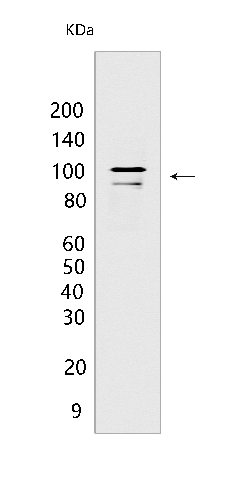
CSE1L Mouse mAb[965B]Cat NO.: A61128
Western blot(SDS PAGE) analysis of extracts from HeLa cells.Using CSE1L Mouse mAb IgG [965B] at dilution of 1:1000 incubated at 4℃ over night.
Product information
Protein names :CSE1L,CAS,XPO2,XPO2_HUMAN,Exportin-2
UniProtID :P55060
MASS(da) :110,417
MW(kDa) :100kDa
Form :Liquid
Purification :Protein A purification
Host :Mouse
Isotype :IgG
sensitivity :Endogenous
Reactivity :Human,Mouse,Rat
- ApplicationDilution
- 免疫印迹(WB)1:1000-2000
- The optimal dilutions should be determined by the end user
Specificity :Antibody is produced by immunizing animals with a synthetic peptide of human CSE1L.
Storage :Antibody store in 10 mM PBS, 0.5mg/ml BSA, 50% glycerol. Shipped at 4°C. Store at-20°C or -80°C. Products are valid for one natural year of receipt.Avoid repeated freeze / thaw cycles.
WB Positive detected :HeLa cells
Function : Export receptor for importin-alpha. Mediates importin-alpha re-export from the nucleus to the cytoplasm after import substrates (cargos) have been released into the nucleoplasm. In the nucleus binds cooperatively to importin-alpha and to the GTPase Ran in its active GTP-bound form. Docking of this trimeric complex to the nuclear pore complex (NPC) is mediated through binding to nucleoporins. Upon transit of a nuclear export complex into the cytoplasm, disassembling of the complex and hydrolysis of Ran-GTP to Ran-GDP (induced by RANBP1 and RANGAP1, respectively) cause release of the importin-alpha from the export receptor. CSE1L/XPO2 then return to the nuclear compartment and mediate another round of transport. The directionality of nuclear export is thought to be conferred by an asymmetric distribution of the GTP- and GDP-bound forms of Ran between the cytoplasm and nucleus..
Tissue specificity :Detected in brain, placenta, ovary, testis and trachea (at protein level) (PubMed:10331944). Widely expressed (PubMed:10331944). Highly expressed in testis and in proliferating cells (PubMed:7479798,PubMed:10331944)..
Subcellular locationi :Cytoplasm. Nucleus.
IMPORTANT: For western blots, incubate membrane with diluted primary antibody in 1% w/v BSA, 1X TBST at 4°C overnight.


