
EPLIN Mouse mAb[5933]Cat NO.: A62578
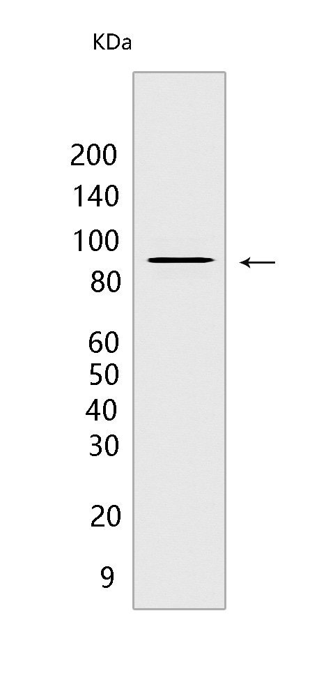
Western blot(SDS PAGE) analysis of extracts from HeLa cells.Using EPLIN Mouse mAb IgG [5933] at dilution of 1:1000 incubated at 4℃ over night.
Product information
Protein names :LIMA1,EPLIN,SREBP3,PP624,LIMA1_HUMAN,LIM domain and actin-binding protein 1
UniProtID :Q9UHB6
MASS(da) :85,226
MW(kDa) :90kDa,110kDa
Form :Liquid
Purification :Protein A purification
Host :Mouse
Isotype :IgG
sensitivity :Endogenous
Reactivity :Human,Rat
- ApplicationDilution
- 免疫印迹(WB)1:1000-2000
- 免疫组化(IHC)1:100
- 免疫荧光(ICC/IF) 1:100,
- The optimal dilutions should be determined by the end user
Specificity :Antibody is produced by immunizing animals with a synthetic peptide of human EPLIN.
Storage :Antibody store in 10 mM PBS, 0.5mg/ml BSA, 50% glycerol. Shipped at 4°C. Store at-20°C or -80°C. Products are valid for one natural year of receipt.Avoid repeated freeze / thaw cycles.
WB Positive detected :HeLa cells
Function : Actin-binding protein involved in actin cytoskeleton regulation and dynamics. Increases the number and size of actin stress fibers and inhibits membrane ruffling. Inhibits actin filament depolymerization. Bundles actin filaments, delays filament nucleation and reduces formation of branched filaments (PubMed:12566430). Plays a role in cholesterol homeostasis. Influences plasma cholesterol levels through regulation of intestinal cholesterol absorption. May act as a scaffold protein by regulating NPC1L1 transportation, an essential protein for cholesterol absorption, to the plasma membrane by recruiting MYO5B to NPC1L1, and thus facilitates cholesterol uptake (By similarity)..
Tissue specificity :Highly expressed in placenta, kidney, pancreas, prostate, ovary, spleen and heart. Also detected in lung, liver, brain, skeletal muscle, thymus, testis and intestine. Not detected in leukocytes. Isoform Beta expressed generally at very low levels. Isoform Alpha abundant in epithelial cells from mammary gland, prostate and in normal oral keratinocytes. Low levels in aortic endothelial cells and dermal fibroblasts. Not detectable in myocardium..
Subcellular locationi :Cytoplasm. Cell junction, focal adhesion. Cytoplasm, cytoskeleton. Cytoplasm, cytoskeleton, stress fiber. Cell membrane.
IMPORTANT: For western blots, incubate membrane with diluted primary antibody in 1% w/v BSA, 1X TBST at 4°C overnight.
-
About
-
Product
-
Law
-
Literature
-

WeChat scan code follow us