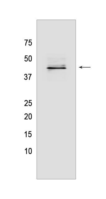
IL-36 Beta/IL-1F8 Mouse mAb[W67F]Cat NO.: A20004
Western blot(SDS PAGE) analysis of extracts from Recombinant protein.Using IL-36 Beta/IL-1F8 Mouse mAb IgG [W67F] at dilution of 1:1000 incubated at 4℃ over night.
Product information
Protein names :IL36B,IL1F8,IL1H2,IL36B_HUMAN,Interleukin-36 beta
UniProtID :Q9NZH7
MASS(da) :18,522
MW(kDa) :44kDa
Form :Liquid
Purification :Protein A purification
Host :Mouse
Isotype :IgG
sensitivity :Endogenous
Reactivity :Human
- ApplicationDilution
- 免疫印迹(WB)1:1000-2000
- 免疫组化(IHC)1:100
- 免疫荧光(ICC/IF) 1:100,
- The optimal dilutions should be determined by the end user
Specificity :Antibody is produced by immunizing animals with a synthetic peptide of human IL-36 Beta/IL-1F8.
Storage :Antibody store in 10 mM PBS, 0.5mg/ml BSA, 50% glycerol. Shipped at 4°C. Store at-20°C or -80°C. Products are valid for one natural year of receipt.Avoid repeated freeze / thaw cycles.
WB Positive detected :Recombinant protein
Function : Cytokine that binds to and signals through the IL1RL2/IL-36R receptor which in turn activates NF-kappa-B and MAPK signaling pathways in target cells linked to a pro-inflammatory response. Part of the IL-36 signaling system that is thought to be present in epithelial barriers and to take part in local inflammatory response,similar to the IL-1 system with which it shares the coreceptor IL1RAP. Stimulates production of interleukin-6 and interleukin-8 in synovial fibrobasts, articular chondrocytes and mature adipocytes. Induces expression of a number of antimicrobial peptides including beta-defensins 4 and 103 as well as a number of matrix metalloproteases. Seems to be involved in skin inflammatory response by acting on keratinocytes, dendritic cells and indirectly on T-cells to drive tissue infiltration, cell maturation and cell proliferation. In cultured keratinocytes induces the expression of macrophage, T-cell, and neutrophil chemokines, such as CCL3, CCL4, CCL5, CCL2, CCL17, CCL22, CL20, CCL5, CCL2, CCL17, CCL22, CXCL8, CCL20 and CXCL1, and the production of pro-inflammatory cytokines such as TNF-alpha, IL-8 and IL-6..
Tissue specificity :Expression at low levels in tonsil, bone marrow, heart, placenta, lung, testis and colon but not in any hematopoietic cell lines. Not detected in adipose tissue. Expressed at higher levels in psoriatic plaques than in symptomless psoriatic skin or healthy control skin. Increased levels are not detected in inflamed joint tissue..
Subcellular locationi :Cytoplasm. Secreted.
IMPORTANT: For western blots, incubate membrane with diluted primary antibody in 1% w/v BSA, 1X TBST at 4°C overnight.


