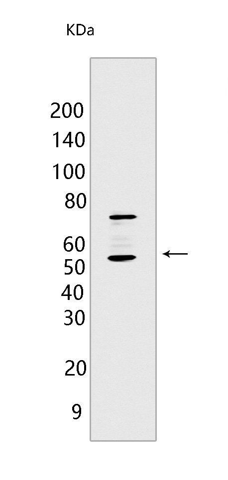
MAVS; VISA Mouse mAb[P5OK]Cat NO.: A51734
Western blot(SDS PAGE) analysis of extracts from A431 cells.Using MAVS; VISA Mouse mAb IgG [P5OK] at dilution of 1:1000 incubated at 4℃ over night.
Product information
Protein names :MAVS,IPS1,KIAA1271,VISA,MAVS_HUMAN,Mitochondrial antiviral-signaling protein
UniProtID :Q7Z434
MASS(da) :56,528
MW(kDa) :70-75kDa,50-55kDa
Form :Liquid
Purification :Protein A purification
Host :Mouse
Isotype :IgG
sensitivity :Endogenous
Reactivity :Human
- ApplicationDilution
- 免疫印迹(WB)1:1000-2000
- 免疫组化(IHC)1:100
- The optimal dilutions should be determined by the end user
Specificity :Antibody is produced by immunizing animals with a synthetic peptide of human MAVS; VISA.
Storage :Antibody store in 10 mM PBS, 0.5mg/ml BSA, 50% glycerol. Shipped at 4°C. Store at-20°C or -80°C. Products are valid for one natural year of receipt.Avoid repeated freeze / thaw cycles.
WB Positive detected :A431 cells
Function : Required for innate immune defense against viruses (PubMed:16125763, PubMed:16127453, PubMed:16153868, PubMed:16177806, PubMed:19631370, PubMed:20451243, PubMed:23087404, PubMed:20127681, PubMed:21170385). Acts downstream of DHX33, DDX58/RIG-I and IFIH1/MDA5, which detect intracellular dsRNA produced during viral replication, to coordinate pathways leading to the activation of NF-kappa-B, IRF3 and IRF7, and to the subsequent induction of antiviral cytokines such as IFNB and RANTES (CCL5) (PubMed:16125763, PubMed:16127453, PubMed:16153868, PubMed:16177806, PubMed:19631370, PubMed:20451243, PubMed:23087404, PubMed:25636800, PubMed:20127681, PubMed:21170385, PubMed:20628368, PubMed:33110251). Peroxisomal and mitochondrial MAVS act sequentially to create an antiviral cellular state (PubMed:20451243). Upon viral infection, peroxisomal MAVS induces the rapid interferon-independent expression of defense factors that provide short-term protection, whereas mitochondrial MAVS activates an interferon-dependent signaling pathway with delayed kinetics, which amplifies and stabilizes the antiviral response (PubMed:20451243). May activate the same pathways following detection of extracellular dsRNA by TLR3 (PubMed:16153868). May protect cells from apoptosis (PubMed:16125763)..
Tissue specificity :Present in T-cells, monocytes, epithelial cells and hepatocytes (at protein level). Ubiquitously expressed, with highest levels in heart, skeletal muscle, liver, placenta and peripheral blood leukocytes..
Subcellular locationi :Mitochondrion outer membrane. Mitochondrion. Peroxisome.
IMPORTANT: For western blots, incubate membrane with diluted primary antibody in 1% w/v BSA, 1X TBST at 4°C overnight.


