
PKC Iota Mouse mAb[7OX4]Cat NO.: A65511
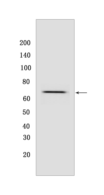
Western blot(SDS PAGE) analysis of extracts from HeLa cells.Using PKC Iota Mouse mAb IgG [7OX4] at dilution of 1:1000 incubated at 4℃ over night.
Product information
Protein names :PRKCI,DXS1179E,KPCI_HUMAN,Protein kinase C iota type
UniProtID :P41743
MASS(da) :68,262
MW(kDa) :70kDa
Form :Liquid
Purification :Protein A purification
Host :Mouse
Isotype :IgG
sensitivity :Endogenous
Reactivity :Human,Mouse,Rat
- ApplicationDilution
- 免疫印迹(WB)1:1000-2000
- 免疫组化(IHC)1:100
- 免疫荧光(ICC/IF) 1:100,
- The optimal dilutions should be determined by the end user
Specificity :Antibody is produced by immunizing animals with a synthetic peptide of human PKC Iota.
Storage :Antibody store in 10 mM PBS, 0.5mg/ml BSA, 50% glycerol. Shipped at 4°C. Store at-20°C or -80°C. Products are valid for one natural year of receipt.Avoid repeated freeze / thaw cycles.
WB Positive detected :HeLa cells
Function : Calcium- and diacylglycerol-independent serine/ threonine-protein kinase that plays a general protective role against apoptotic stimuli, is involved in NF-kappa-B activation, cell survival, differentiation and polarity, and contributes to the regulation of microtubule dynamics in the early secretory pathway. Is necessary for BCR-ABL oncogene-mediated resistance to apoptotic drug in leukemia cells, protecting leukemia cells against drug-induced apoptosis. In cultured neurons, prevents amyloid beta protein-induced apoptosis by interrupting cell death process at a very early step. In glioblastoma cells, may function downstream of phosphatidylinositol 3-kinase (PI(3)K) and PDPK1 in the promotion of cell survival by phosphorylating and inhibiting the pro-apoptotic factor BAD. Can form a protein complex in non-small cell lung cancer (NSCLC) cells with PARD6A and ECT2 and regulate ECT2 oncogenic activity by phosphorylation, which in turn promotes transformed growth and invasion. In response to nerve growth factor (NGF), acts downstream of SRC to phosphorylate and activate IRAK1, allowing the subsequent activation of NF-kappa-B and neuronal cell survival. Functions in the organization of the apical domain in epithelial cells by phosphorylating EZR. This step is crucial for activation and normal distribution of EZR at the early stages of intestinal epithelial cell differentiation. Forms a protein complex with LLGL1 and PARD6B independently of PARD3 to regulate epithelial cell polarity. Plays a role in microtubule dynamics in the early secretory pathway through interaction with RAB2A and GAPDH and recruitment to vesicular tubular clusters (VTCs). In human coronary artery endothelial cells (HCAEC), is activated by saturated fatty acids and mediates lipid-induced apoptosis. Involved in early synaptic long term potentiation phase in CA1 hippocampal cells and short term memory formation (By similarity)..
Tissue specificity :Predominantly expressed in lung and brain, but also expressed at lower levels in many tissues including pancreatic islets. Highly expressed in non-small cell lung cancers..
Subcellular locationi :Cytoplasm. Membrane. Endosome. Nucleus.
IMPORTANT: For western blots, incubate membrane with diluted primary antibody in 1% w/v BSA, 1X TBST at 4°C overnight.
-
About
-
Product
-
Law
-
Literature
-

WeChat scan code follow us