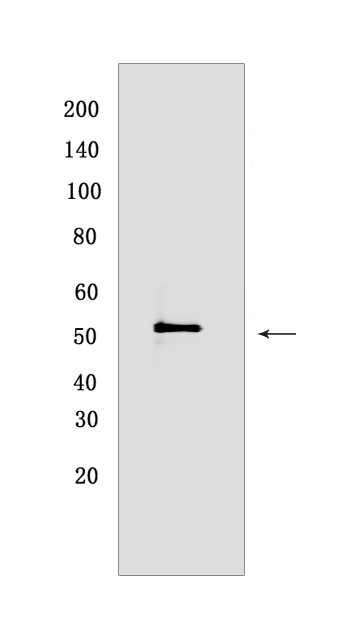
VISTA Rabbit mAb [6L4J]Cat NO.: A65955
Western blot(SDS PAGE) analysis of extracts from MCF-7 cells.Using VISTA Rabbit mAb [6L4J] at dilution of 1:1000 incubated at 4℃ over night.
Product information
Protein names :VSIR,C10orf54,SISP1,VISTA,PP2135,UNQ730/PRO1412,VISTA_HUMAN,V-type immunoglobulin domain-containing suppressor of T-cell activation
UniProtID :Q9H7M9
MW(kDa) :45-65 kDa
Form :Liquid
Purification :Protein A purification
Host :Rabbit
Isotype :IgG
sensitivity :Endogenous
Reactivity :Human
- ApplicationDilution
- 免疫印迹(WB)1:1000-2000
- The optimal dilutions should be determined by the end user
Specificity :Antibody is produced by immunizing animals with a synthetic peptide at the sequence of Human VISTA
Storage :Antibody store in 10 mM PBS, 0.5mg/ml BSA, 50% glycerol. Shipped at 4°C. Store at-20°C or -80°C. Products are valid for one natural year of receipt.Avoid repeated freeze / thaw cycles.
WB Positive detected :MCF-7 cells
Function : Immunoregulatory receptor which inhibits the T-cell response (PubMed:24691993). May promote differentiation of embryonic stem cells, by inhibiting BMP4 signaling (By similarity). May stimulate MMP14-mediated MMP2 activation (PubMed:20666777)..
Tissue specificity :Expressed in spleen. Detected on a number of myeloid cells including CD11b monocytes, CD66b+ neutrophils, at low levels on CD4+ and CD8+ T-cells, and in a subset of NK cells. Not detected on B cells (at protein level). Expressed at high levels in placenta, spleen, plasma blood leukocytes, and lung. Expressed at moderate levels in lymph node, bone marrow, fat, uterus, and trachea. Has low expression levels in other tissues..
Subcellular locationi :Cell membrane,Single-pass type I membrane protein.
IMPORTANT: For western blots, incubate membrane with diluted primary antibody in 1% w/v BSA, 1X TBST at 4°C overnight.


