
TNFR1 Mouse mAb[OIRT]Cat NO.: A21246
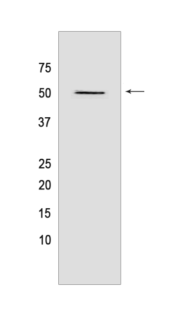
Western blot(SDS PAGE) analysis of extracts from HL-60 cells.Using TNFR1 Mouse mAb IgG [OIRT] at dilution of 1:1000 incubated at 4℃ over night.
Product information
Protein names :TNFRSF1A,TNFAR,TNFR1,TNR1A_HUMAN,Tumor necrosis factor receptor superfamily member 1A [Cleaved into: Tumor necrosis factor receptor superfamily member 1A, membrane form,Tumor necrosis factor-binding protein 1 ]
UniProtID :P19438
MASS(da) :50,495
MW(kDa) :50kda
Form :Liquid
Purification :Protein A purification
Host :Mouse
Isotype :IgG
sensitivity :Endogenous
Reactivity :Human,Mouse,Rat
- ApplicationDilution
- 免疫印迹(WB)1:1000-2000
- 免疫组化(IHC)1:100
- 免疫荧光(ICC/IF) 1:100,
- The optimal dilutions should be determined by the end user
Specificity :Antibody is produced by immunizing animals with a synthetic peptide of human TNFR1.
Storage :Antibody store in 10 mM PBS, 0.5mg/ml BSA, 50% glycerol. Shipped at 4°C. Store at-20°C or -80°C. Products are valid for one natural year of receipt.Avoid repeated freeze / thaw cycles.
WB Positive detected :HL-60 cells
Function : Receptor for TNFSF2/TNF-alpha and homotrimeric TNFSF1/lymphotoxin-alpha. The adapter molecule FADD recruits caspase-8 to the activated receptor. The resulting death-inducing signaling complex (DISC) performs caspase-8 proteolytic activation which initiates the subsequent cascade of caspases (aspartate-specific cysteine proteases) mediating apoptosis. Contributes to the induction of non-cytocidal TNF effects including anti-viral state and activation of the acid sphingomyelinase.
Subcellular locationi :Cell membrane,Single-pass type I membrane protein. Golgi apparatus membrane,Single-pass type I membrane protein. Secreted.
IMPORTANT: For western blots, incubate membrane with diluted primary antibody in 1% w/v BSA, 1X TBST at 4°C overnight.
-
About
-
Product
-
Law
-
Literature
-

WeChat scan code follow us