
DR6 Mouse mAb[55Z4]Cat NO.: A37234
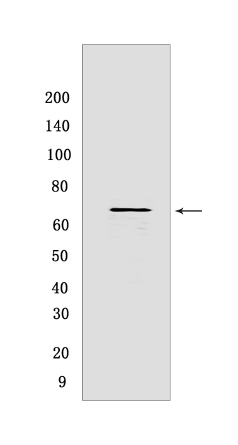
Western blot(SDS PAGE) analysis of extracts from K-562 cells.Using DR6 Mouse mAb IgG [55Z4] at dilution of 1:1000 incubated at 4℃ over night.
Product information
Protein names :TNFRSF21,DR6,UNQ437/PRO868,TNR21_HUMAN,Tumor necrosis factor receptor superfamily member 21
UniProtID :O75509
MASS(da) :71,845
MW(kDa) :69kDa
Form :Liquid
Purification :Protein A purification
Host :Mouse
Isotype :IgG
sensitivity :Endogenous
Reactivity :Human
- ApplicationDilution
- 免疫印迹(WB)1:1000-2000
- The optimal dilutions should be determined by the end user
Specificity :Antibody is produced by immunizing animals with a synthetic peptide of human DR6.
Storage :Antibody store in 10 mM PBS, 0.5mg/ml BSA, 50% glycerol. Shipped at 4°C. Store at-20°C or -80°C. Products are valid for one natural year of receipt.Avoid repeated freeze / thaw cycles.
WB Positive detected :K-562 cells
Function : Promotes apoptosis, possibly via a pathway that involves the activation of NF-kappa-B. Can also promote apoptosis mediated by BAX and by the release of cytochrome c from the mitochondria into the cytoplasm. Plays a role in neuronal apoptosis, including apoptosis in response to amyloid peptides derived from APP, and is required for both normal cell body death and axonal pruning. Trophic-factor deprivation triggers the cleavage of surface APP by beta-secretase to release sAPP-beta which is further cleaved to release an N-terminal fragment of APP (N-APP). N-APP binds TNFRSF21,this triggers caspase activation and degeneration of both neuronal cell bodies (via caspase-3) and axons (via caspase-6). Negatively regulates oligodendrocyte survival, maturation and myelination. Plays a role in signaling cascades triggered by stimulation of T-cell receptors, in the adaptive immune response and in the regulation of T-cell differentiation and proliferation. Negatively regulates T-cell responses and the release of cytokines such as IL4, IL5, IL10, IL13 and IFNG by Th2 cells. Negatively regulates the production of IgG, IgM and IgM in response to antigens. May inhibit the activation of JNK in response to T-cell stimulation..
Tissue specificity :Detected in fetal spinal cord and in brain neurons, with higher levels in brain from Alzheimer disease patients (at protein level). Highly expressed in heart, brain, placenta, pancreas, lymph node, thymus and prostate. Detected at lower levels in lung, skeletal muscle, kidney, testis, uterus, small intestine, colon, spleen, bone marrow and fetal liver. Very low levels were found in adult liver and peripheral blood leukocytes..
Subcellular locationi :Cell membrane,Single-pass type I membrane protein.
IMPORTANT: For western blots, incubate membrane with diluted primary antibody in 1% w/v BSA, 1X TBST at 4°C overnight.
-
About
-
Product
-
Law
-
Literature
-

WeChat scan code follow us