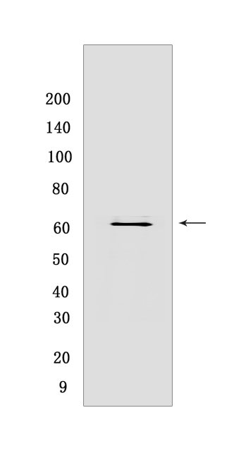
IFIT3 Mouse mAb[03IY]Cat NO.: A69895
Western blot(SDS PAGE) analysis of extracts from HeLa cells.Using IFIT3 Mouse mAb IgG [03IY] at dilution of 1:1000 incubated at 4℃ over night.
Product information
Protein names :IFIT3,CIG-49,IFI60,IFIT4,ISG60,IFIT3_HUMAN,Interferon-induced protein with tetratricopeptide repeats 3
UniProtID :O14879
MASS(da) :55,985
MW(kDa) :60kDa
Form :Liquid
Purification :Protein A purification
Host :Mouse
Isotype :IgG
sensitivity :Endogenous
Reactivity :Human
- ApplicationDilution
- 免疫印迹(WB)1:1000-2000
- 免疫组化(IHC)1:100
- 免疫荧光(ICC/IF) 1:100,
- The optimal dilutions should be determined by the end user
Specificity :Antibody is produced by immunizing animals with a synthetic peptide of human IFIT3.
Storage :Antibody store in 10 mM PBS, 0.5mg/ml BSA, 50% glycerol. Shipped at 4°C. Store at-20°C or -80°C. Products are valid for one natural year of receipt.Avoid repeated freeze / thaw cycles.
WB Positive detected :HeLa cells
Function : IFN-induced antiviral protein which acts as an inhibitor of cellular as well as viral processes, cell migration, proliferation, signaling, and viral replication. Enhances MAVS-mediated host antiviral responses by serving as an adapter bridging TBK1 to MAVS which leads to the activation of TBK1 and phosphorylation of IRF3 and phosphorylated IRF3 translocates into nucleus to promote antiviral gene transcription. Exhibits an antiproliferative activity via the up-regulation of cell cycle negative regulators CDKN1A/p21 and CDKN1B/p27. Normally, CDKN1B/p27 turnover is regulated by COPS5, which binds CDKN1B/p27 in the nucleus and exports it to the cytoplasm for ubiquitin-dependent degradation. IFIT3 sequesters COPS5 in the cytoplasm, thereby increasing nuclear CDKN1B/p27 protein levels. Up-regulates CDKN1A/p21 by down-regulating MYC, a repressor of CDKN1A/p21. Can negatively regulate the apoptotic effects of IFIT2..
Tissue specificity :Expression significantly higher in peripheral blood mononuclear cells (PBMCs) and monocytes from systemic lupus erythematosus (SLE) patients than in those from healthy individuals (at protein level). Spleen, lung, leukocytes, lymph nodes, placenta, bone marrow and fetal liver..
Subcellular locationi :Cytoplasm. Mitochondrion.
IMPORTANT: For western blots, incubate membrane with diluted primary antibody in 1% w/v BSA, 1X TBST at 4°C overnight.


