
Erk5 Rabbit mAb [1WI9]Cat NO.: A75491
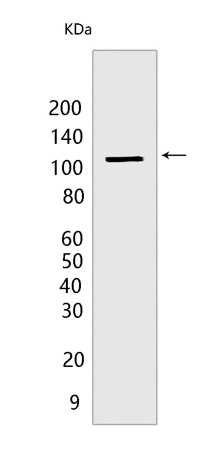
Western blot(SDS PAGE) analysis of extracts from COS-7 cells.Using Erk5 Rabbit mAb [1WI9] at dilution of 1:1000 incubated at 4℃ over night.
Product information
Protein names :MAPK7,BMK1,ERK5,PRKM7,MK07_HUMAN,Mitogen-activated protein kinase 7
UniProtID :Q13164
MASS(da) :88,386
MW(kDa) :115 kDa
Form :Liquid
Purification :Protein A purification
Host :Rabbit
Isotype :IgG
sensitivity :Endogenous
Reactivity :Human,Mouse,Rat
- ApplicationDilution
- 免疫印迹(WB)1:1000-2000
- The optimal dilutions should be determined by the end user
Specificity :Antibody is produced by immunizing animals with a synthetic peptide at the sequence of Human Erk5
Storage :Antibody store in 10 mM PBS, 0.5mg/ml BSA, 50% glycerol. Shipped at 4°C. Store at-20°C or -80°C. Products are valid for one natural year of receipt.Avoid repeated freeze / thaw cycles.
WB Positive detected :COS-7 cells
Function : Plays a role in various cellular processes such as proliferation, differentiation and cell survival. The upstream activator of MAPK7 is the MAPK kinase MAP2K5. Upon activation, it translocates to the nucleus and phosphorylates various downstream targets including MEF2C. EGF activates MAPK7 through a Ras-independent and MAP2K5-dependent pathway. May have a role in muscle cell differentiation. May be important for endothelial function and maintenance of blood vessel integrity. MAP2K5 and MAPK7 interact specifically with one another and not with MEK1/ERK1 or MEK2/ERK2 pathways. Phosphorylates SGK1 at Ser-78 and this is required for growth factor-induced cell cycle progression. Involved in the regulation of p53/TP53 by disrupting the PML-MDM2 interaction..
Tissue specificity :Expressed in many adult tissues. Abundant in heart, placenta, lung, kidney and skeletal muscle. Not detectable in liver..
Subcellular locationi :Cytoplasm. Nucleus. Nucleus, PML body.
IMPORTANT: For western blots, incubate membrane with diluted primary antibody in 1% w/v BSA, 1X TBST at 4°C overnight.
-
About
-
Product
-
Law
-
Literature
-

WeChat scan code follow us