
KIDINS220 Mouse mAb[4W95]Cat NO.: A90870
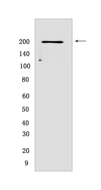
Western blot(SDS PAGE) analysis of extracts from HEK-293 cells.Using KIDINS220 Mouse mAb IgG [4W95] at dilution of 1:1000 incubated at 4℃ over night.
Product information
Protein names :KIDINS220,ARMS,KIAA1250,KDIS_HUMAN,Kinase D-interacting substrate of 220 kDa
UniProtID :Q9ULH0
MASS(da) :196,542
MW(kDa) :200kDa
Form :Liquid
Purification :Protein A purification
Host :Mouse
Isotype :IgG
sensitivity :Endogenous
Reactivity :Human
- ApplicationDilution
- 免疫印迹(WB)1:1000-2000
- The optimal dilutions should be determined by the end user
Specificity :Antibody is produced by immunizing animals with a synthetic peptide of human KIDINS220.
Storage :Antibody store in 10 mM PBS, 0.5mg/ml BSA, 50% glycerol. Shipped at 4°C. Store at-20°C or -80°C. Products are valid for one natural year of receipt.Avoid repeated freeze / thaw cycles.
WB Positive detected :HEK-293 cells
Function : Promotes a prolonged MAP-kinase signaling by neurotrophins through activation of a Rap1-dependent mechanism. Provides a docking site for the CRKL-C3G complex, resulting in Rap1-dependent sustained ERK activation. May play an important role in regulating postsynaptic signal transduction through the syntrophin-mediated localization of receptor tyrosine kinases such as EPHA4. In cooperation with SNTA1 can enhance EPHA4-induced JAK/STAT activation. Plays a role in nerve growth factor (NGF)-induced recruitment of RAPGEF2 to late endosomes and neurite outgrowth. May play a role in neurotrophin- and ephrin-mediated neuronal outgrowth and in axon guidance during neural development and in neuronal regeneration (By similarity). Modulates stress-induced apoptosis of melanoma cells via regulation of the MEK/ERK signaling pathway..
Tissue specificity :Abundant in developing and adult neural tissues as well as neuroendocrine cells and dendritic cells. Overexpressed in melanoma and melanoma cell lines..
Subcellular locationi :Membrane,Multi-pass membrane protein. Late endosome.
IMPORTANT: For western blots, incubate membrane with diluted primary antibody in 1% w/v BSA, 1X TBST at 4°C overnight.
-
About
-
Product
-
Law
-
Literature
-

WeChat scan code follow us