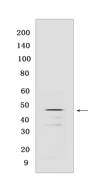
P-GSK-3-beta (S9) Rabbit mAb [46J0]Cat NO.: A76960
Western blot(SDS PAGE) analysis of extracts from NIH/3T3 cells PDGF-treated (100 μg/ml 15 min).Using P-GSK-3-beta (S9) Rabbit mAb [46J0] at dilution of 1:1000 incubated at 4℃ over night.
Product information
Protein names :GSK3B,GSK3B_HUMAN,Glycogen synthase kinase-3 beta
UniProtID :P49841
MASS(da) :46,744
MW(kDa) :46 kDa
Form :Liquid
Purification :Protein A purification
Host :Rabbit
Isotype :IgG
sensitivity :Endogenous
Reactivity :Human,Mouse,Rat
- ApplicationDilution
- 免疫印迹(WB)1:1000-2000
- The optimal dilutions should be determined by the end user
Specificity :Antibody is produced by immunizing animals with a synthetic peptide at the sequence of Human Phospho-GSK-3-beta (Ser9)
Storage :Antibody store in 10 mM PBS, 0.5mg/ml BSA, 50% glycerol. Shipped at 4°C. Store at-20°C or -80°C. Products are valid for one natural year of receipt.Avoid repeated freeze / thaw cycles.
WB Positive detected :NIH/3T3 cells PDGF-treated (100 μg/ml 15 min)
Function : Constitutively active protein kinase that acts as a negative regulator in the hormonal control of glucose homeostasis, Wnt signaling and regulation of transcription factors and microtubules, by phosphorylating and inactivating glycogen synthase (GYS1 or GYS2), EIF2B, CTNNB1/beta-catenin, APC, AXIN1, DPYSL2/CRMP2, JUN, NFATC1/NFATC, MAPT/TAU and MACF1 (PubMed:1846781, PubMed:9072970, PubMed:14690523, PubMed:20937854, PubMed:12554650, PubMed:11430833, PubMed:16484495). Requires primed phosphorylation of the majority of its substrates (PubMed:11430833, PubMed:16484495). In skeletal muscle, contributes to insulin regulation of glycogen synthesis by phosphorylating and inhibiting GYS1 activity and hence glycogen synthesis (PubMed:8397507). May also mediate the development of insulin resistance by regulating activation of transcription factors (PubMed:8397507). Regulates protein synthesis by controlling the activity of initiation factor 2B (EIF2BE/EIF2B5) in the same manner as glycogen synthase (PubMed:8397507). In Wnt signaling, GSK3B forms a multimeric complex with APC, AXIN1 and CTNNB1/beta-catenin and phosphorylates the N-terminus of CTNNB1 leading to its degradation mediated by ubiquitin/proteasomes (PubMed:12554650). Phosphorylates JUN at sites proximal to its DNA-binding domain, thereby reducing its affinity for DNA (PubMed:1846781). Phosphorylates NFATC1/NFATC on conserved serine residues promoting NFATC1/NFATC nuclear export, shutting off NFATC1/NFATC gene regulation, and thereby opposing the action of calcineurin (PubMed:9072970). Phosphorylates MAPT/TAU on 'Thr-548', decreasing significantly MAPT/TAU ability to bind and stabilize microtubules (PubMed:14690523). MAPT/TAU is the principal component of neurofibrillary tangles in Alzheimer disease (PubMed:14690523). Plays an important role in ERBB2-dependent stabilization of microtubules at the cell cortex (PubMed:20937854). Phosphorylates MACF1, inhibiting its binding to microtubules which is critical for its role in bulge stem cell migration and skin wound repair (By similarity). Probably regulates NF-kappa-B (NFKB1) at the transcriptional level and is required for the NF-kappa-B-mediated anti-apoptotic response to TNF-alpha (TNF/TNFA) (By similarity). Negatively regulates replication in pancreatic beta-cells, resulting in apoptosis, loss of beta-cells and diabetes (By similarity). Through phosphorylation of the anti-apoptotic protein MCL1, may control cell apoptosis in response to growth factors deprivation (By similarity). Phosphorylates MUC1 in breast cancer cells, decreasing the interaction of MUC1 with CTNNB1/beta-catenin (PubMed:9819408). Is necessary for the establishment of neuronal polarity and axon outgrowth (PubMed:20067585). Phosphorylates MARK2, leading to inhibition of its activity (By similarity). Phosphorylates SIK1 at 'Thr-182', leading to sustainment of its activity (PubMed:18348280). Phosphorylates ZC3HAV1 which enhances its antiviral activity (PubMed:22514281). Phosphorylates SNAI1, leading to its BTRC-triggered ubiquitination and proteasomal degradation (PubMed:15448698, PubMed:15647282). Phosphorylates SFPQ at 'Thr-687' upon T-cell activation (PubMed:20932480). Phosphorylates NR1D1 st 'Ser-55' and 'Ser-59' and stabilizes it by protecting it from proteasomal degradation. Regulates the circadian clock via phosphorylation of the major clock components including ARNTL/BMAL1, CLOCK and PER2 (PubMed:19946213, PubMed:28903391). Phosphorylates CLOCK AT 'Ser-427' and targets it for proteasomal degradation (PubMed:19946213). Phosphorylates ARNTL/BMAL1 at 'Ser-17' and 'Ser-21' and primes it for ubiquitination and proteasomal degradation (PubMed:28903391). Phosphorylates OGT at 'Ser-3' or 'Ser-4' which positively regulates its activity. Phosphorylates MYCN in neuroblastoma cells which may promote its degradation (PubMed:24391509). Regulates the circadian rhythmicity of hippocampal long-term potentiation and ARNTL/BMLA1 and PER2 expression (By similarity). Acts as a regulator of autophagy by mediating phosphorylation of KAT5/TIP60 under starvation conditions, activating KAT5/TIP60 acetyltransferase activity and promoting acetylation of key autophagy regulators, such as ULK1 and RUBCNL/Pacer (PubMed:30704899). Negatively regulates extrinsic apoptotic signaling pathway via death domain receptors. Promotes the formation of an anti-apoptotic complex, made of DDX3X, BRIC2 and GSK3B, at death receptors, including TNFRSF10B. The anti-apoptotic function is most effective with weak apoptotic signals and can be overcome by stronger stimulation (PubMed:18846110). Phosphorylates E2F1, promoting the interaction between E2F1 and USP11, stabilizing E2F1 and promoting its activity (PubMed:17050006, PubMed:28992046). Phosphorylates mTORC2 complex component RICTOR at 'Thr-1695' which facilitates FBXW7-mediated ubiquitination and subsequent degradation of RICTOR (PubMed:25897075)..
Tissue specificity :Expressed in testis, thymus, prostate and ovary and weakly expressed in lung, brain and kidney. Colocalizes with EIF2AK2/PKR and TAU in the Alzheimer disease (AD) brain..
Subcellular locationi :Cytoplasm. Nucleus. Cell membrane.
IMPORTANT: For western blots, incubate membrane with diluted primary antibody in 1% w/v BSA, 1X TBST at 4°C overnight.


