
TFDP1 Rabbit mAb [CQV0]Cat NO.: A40177
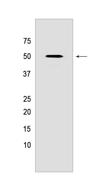
Western blot(SDS PAGE) analysis of extracts from HeLa cells.Using TFDP1 Rabbit mAb [CQV0] at dilution of 1:1000 incubated at 4℃ over night.
Product information
Protein names :TFDP1,DP1,TFDP1_HUMAN,Transcription factor Dp-1
UniProtID :Q14186
MASS(da) :45,070
MW(kDa) :50KDa
Form :Liquid
Purification :Protein A purification
Host :Rabbit monoclonal IgG
Isotype :IgG
sensitivity :Endogenous
Reactivity :Human,Rat
- ApplicationDilution
- 免疫印迹(WB)1:1000-2000
- 免疫组化(IHC)1:100
- The optimal dilutions should be determined by the end user
Specificity :Antibody is produced by immunizing animals with a synthetic peptide of human TFDP1.
Storage :Antibody store in 10 mM PBS, 0.5mg/ml BSA, 50% glycerol. Shipped at 4°C. Store at-20°C or -80°C. Products are valid for one natural year of receipt.Avoid repeated freeze / thaw cycles.
WB Positive detected :HeLa cells
Function : Can stimulate E2F-dependent transcription. Binds DNA cooperatively with E2F family members through the E2 recognition site, 5'-TTTC[CG]CGC-3', found in the promoter region of a number of genes whose products are involved in cell cycle regulation or in DNA replication (PubMed:8405995, PubMed:7739537). The E2F1:DP complex appears to mediate both cell proliferation and apoptosis. Blocks adipocyte differentiation by repressing CEBPA binding to its target gene promoters (PubMed:20176812)..
Tissue specificity :Highest levels in muscle. Also expressed in brain, placenta, liver and kidney. Lower levels in lung and pancreas. Not detected in heart.
Subcellular locationi :Nucleus. Cytoplasm.
IMPORTANT: For western blots, incubate membrane with diluted primary antibody in 1% w/v BSA, 1X TBST at 4°C overnight.
-
About
-
Product
-
Law
-
Literature
-

WeChat scan code follow us