
Rhodopsin Rabbit mAb [G580]Cat NO.: A37793
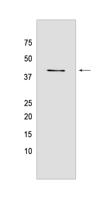
Western blot analysis of extracts from Rat eyeball tissue lyaste.using Rhodopsin Rabbit mAb [G580] at dilution of 1:1000 incubated at 4℃ over night
Product information
Protein names :RHO,OPN2,OPSD_HUMAN,Rhodopsin
UniProtID :P08100
MASS(da) :38,893
MW(kDa) :39 kDa
Form :Liquid
Purification :Protein A purification
Host :Rabbit
Isotype :IgG
sensitivity :Endogenous
Reactivity :Human,Rat
- ApplicationDilution
- 免疫印迹(WB)1:1000-2000
- 免疫组化(IHC)1:100,
- The optimal dilutions should be determined by the end user
Specificity :Antibody is produced by immunizing animals with a synthetic peptide of Human Rhodopsin.
Storage :Antibody store in 10 mM PBS, 0.5mg/ml BSA, 50% glycerol. Shipped at 4°C. Store at-20°C or -80°C. Products are valid for one natural year of receipt.Avoid repeated freeze / thaw cycles.
WB Positive detected :Rat eyeball tissue lyaste
Function : Photoreceptor required for image-forming vision at low light intensity (PubMed:8107847, PubMed:7846071). Required for photoreceptor cell viability after birth (PubMed:2215617, PubMed:12566452). Light-induced isomerization of the chromophore 11-cis-retinal to all-trans-retinal triggers a conformational change that activates signaling via G-proteins (PubMed:8107847, PubMed:28524165, PubMed:26200343, PubMed:28753425). Subsequent receptor phosphorylation mediates displacement of the bound G-protein alpha subunit by the arrestin SAG and terminates signaling (PubMed:28524165, PubMed:26200343)..
Tissue specificity :Rod shaped photoreceptor cells which mediate vision in dim light.
Subcellular locationi :Membrane,Multi-pass membrane protein. Cell projection, cilium, photoreceptor outer segment.
IMPORTANT: For western blots, incubate membrane with diluted primary antibody in 1% w/v BSA, 1X TBST at 4°C overnight.
-
About
-
Product
-
Law
-
Literature
-

WeChat scan code follow us