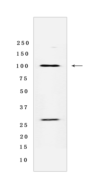
Melanoma gp100 Rabbit mAb [12XB]Cat NO.: A34015
Western blot analysis of extracts from Human melanoma tissue lyaste.using Melanoma gp100 Rabbit mAb [12XB] at dilution of 1:1000 incubated at 4℃ over night
Product information
Protein names :PMEL,D12S53E,PMEL17,SILV,PMEL_HUMAN,Melanocyte protein PMEL [Cleaved into: M-alpha ,M-beta]
UniProtID :P40967
MASS(da) :70,255
MW(kDa) :100, 26 kDa
Form :Liquid
Purification :Protein A purification
Host :Rabbit
Isotype :IgG
sensitivity :Endogenous
Reactivity :Human
- ApplicationDilution
- 免疫印迹(WB)1:1000-2000
- 免疫组化(IHC)1:100,
- The optimal dilutions should be determined by the end user
Specificity :Antibody is produced by immunizing animals with a synthetic peptide of Human Melanoma gp100.
Storage :Antibody store in 10 mM PBS, 0.5mg/ml BSA, 50% glycerol. Shipped at 4°C. Store at-20°C or -80°C. Products are valid for one natural year of receipt.Avoid repeated freeze / thaw cycles.
WB Positive detected :Human melanoma tissue lyaste
Function : Plays a central role in the biogenesis of melanosomes. Involved in the maturation of melanosomes from stage I to II. The transition from stage I melanosomes to stage II melanosomes involves an elongation of the vesicle, and the appearance within of distinct fibrillar structures. Release of the soluble form, ME20-S, could protect tumor cells from antibody mediated immunity..
Tissue specificity :Preferentially expressed in melanomas. Some expression was found in dysplastic nevi. Not found in normal tissues nor in carcinomas. Normally expressed at low levels in quiescent adult melanocytes but overexpressed by proliferating neonatal melanocytes and during tumor growth.
Subcellular locationi :Endoplasmic reticulum membrane,Single-pass type I membrane protein. Golgi apparatus. Melanosome. Endosome, multivesicular body.
IMPORTANT: For western blots, incubate membrane with diluted primary antibody in 1% w/v BSA, 1X TBST at 4°C overnight.


