
HLA-DRA Rabbit mAb [3W29]Cat NO.: A23661
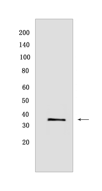
Western blot(SDS PAGE) analysis of extracts from Raji cells.Using HLA-DRA Rabbit mAb [3W29] at dilution of 1:1000 incubated at 4℃ over night.
Product information
Protein names :HLA-DRA,HLA-DRA1,DRA_HUMAN,HLA class II histocompatibility antigen, DR alpha chain
UniProtID :P01903
MASS(da) :28,621
MW(kDa) :30-40 kDa
Form :Liquid
Purification :Protein A purification
Host :Rabbit
Isotype :IgG
sensitivity :Endogenous
Reactivity :Human
- ApplicationDilution
- 免疫印迹(WB)1:1000-2000
- The optimal dilutions should be determined by the end user
Specificity :Antibody is produced by immunizing animals with a synthetic peptide at the sequence of Human HLA-DRA
Storage :Antibody store in 10 mM PBS, 0.5mg/ml BSA, 50% glycerol. Shipped at 4°C. Store at-20°C or -80°C. Products are valid for one natural year of receipt.Avoid repeated freeze / thaw cycles.
WB Positive detected :Raji cells
Function : An alpha chain of antigen-presenting major histocompatibility complex class II (MHCII) molecule. In complex with the beta chain HLA-DRB, displays antigenic peptides on professional antigen presenting cells (APCs) for recognition by alpha-beta T cell receptor (TCR) on HLA-DR-restricted CD4-positive T cells. This guides antigen-specific T-helper effector functions, both antibody-mediated immune response and macrophage activation, to ultimately eliminate the infectious agents and transformed cells (PubMed:29884618, PubMed:17334368, PubMed:8145819, PubMed:15322540, PubMed:22327072, PubMed:27591323, PubMed:31495665, PubMed:15265931, PubMed:9075930, PubMed:24190431). Typically presents extracellular peptide antigens of 10 to 30 amino acids that arise from proteolysis of endocytosed antigens in lysosomes (PubMed:8145819). In the tumor microenvironment, presents antigenic peptides that are primarily generated in tumor-resident APCs likely via phagocytosis of apoptotic tumor cells or macropinocytosis of secreted tumor proteins (PubMed:31495665). Presents peptides derived from intracellular proteins that are trapped in autolysosomes after macroautophagy, a mechanism especially relevant for T cell selection in the thymus and central immune tolerance (PubMed:17182262, PubMed:23783831). The selection of the immunodominant epitopes follows two processing modes: 'bind first, cut/trim later' for pathogen-derived antigenic peptides and 'cut first, bind later' for autoantigens/self-peptides (PubMed:25413013). The anchor residue at position 1 of the peptide N-terminus, usually a large hydrophobic residue, is essential for high affinity interaction with MHCII molecules (PubMed:8145819)..
Tissue specificity :Expressed in professional APCs: macrophages, dendritic cells and B cells (at protein level) (PubMed:31495665, PubMed:15322540, PubMed:23783831). Expressed in thymic epithelial cells (at protein level) (PubMed:23783831)..
Subcellular locationi :Cell membrane,Single-pass type I membrane protein. Endoplasmic reticulum membrane,Single-pass type I membrane protein. Early endosome membrane,Single-pass type I membrane protein. Late endosome membrane,Single-pass type I membrane protein. Lysosome membrane,Single-pass type I membrane protein. Autolysosome membrane,Single-pass type I membrane protein.
IMPORTANT: For western blots, incubate membrane with diluted primary antibody in 1% w/v BSA, 1X TBST at 4°C overnight.
-
About
-
Product
-
Law
-
Literature
-

WeChat scan code follow us