JNK3 Rabbit mAb [B025]Cat NO.: A26067
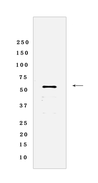
Western blot analysis of extracts from HeLa cells lyastes.using JNK3 Rabbit mAb [B025] at dilution of 1:1000 incubated at 4℃ over night
Product information
Protein names :MAPK10,JNK3,JNK3A,PRKM10,SAPK1B,MK10_HUMAN,Mitogen-activated protein kinase 10
UniProtID :P53779
MASS(da) :52,585
MW(kDa) :53 kDa
Form :Liquid
Purification :Protein A purification
Host :Rabbit
Isotype :IgG
sensitivity :Endogenous
Reactivity :Human
- ApplicationDilution
- 免疫印迹(WB)1:1000-2000
- 免疫荧光(ICC/IF)1:100,
- The optimal dilutions should be determined by the end user
Specificity :Antibody is produced by immunizing animals with a synthetic peptide of Human JNK3.
Storage :Antibody store in 10 mM PBS, 0.5mg/ml BSA, 50% glycerol. Shipped at 4°C. Store at-20°C or -80°C. Products are valid for one natural year of receipt.Avoid repeated freeze / thaw cycles.
WB Positive detected :HeLa cells lyastes
Function : Serine/threonine-protein kinase involved in various processes such as neuronal proliferation, differentiation, migration and programmed cell death. Extracellular stimuli such as pro-inflammatory cytokines or physical stress stimulate the stress-activated protein kinase/c-Jun N-terminal kinase (SAP/JNK) signaling pathway. In this cascade, two dual specificity kinases MAP2K4/MKK4 and MAP2K7/MKK7 phosphorylate and activate MAPK10/JNK3. In turn, MAPK10/JNK3 phosphorylates a number of transcription factors, primarily components of AP-1 such as JUN and ATF2 and thus regulates AP-1 transcriptional activity. Plays regulatory roles in the signaling pathways during neuronal apoptosis. Phosphorylates the neuronal microtubule regulator STMN2. Acts in the regulation of the amyloid-beta precursor protein/APP signaling during neuronal differentiation by phosphorylating APP. Participates also in neurite growth in spiral ganglion neurons. Phosphorylates the CLOCK-ARNTL/BMAL1 heterodimer and plays a role in the photic regulation of the circadian clock (PubMed:22441692). Phosphorylates JUND and this phosphorylation is inhibited in the presence of MEN1 (PubMed:22327296)..
Tissue specificity :Specific to a subset of neurons in the nervous system. Present in the hippocampus and areas, cerebellum, striatum, brain stem, and weakly in the spinal cord. Very weak expression in testis and kidney.
Subcellular locationi :Cytoplasm. Membrane,Lipid-anchor. Nucleus. Mitochondrion.
IMPORTANT: For western blots, incubate membrane with diluted primary antibody in 1% w/v BSA, 1X TBST at 4°C overnight.


