LAG3 Rabbit mAb [M71Y]Cat NO.: A94912
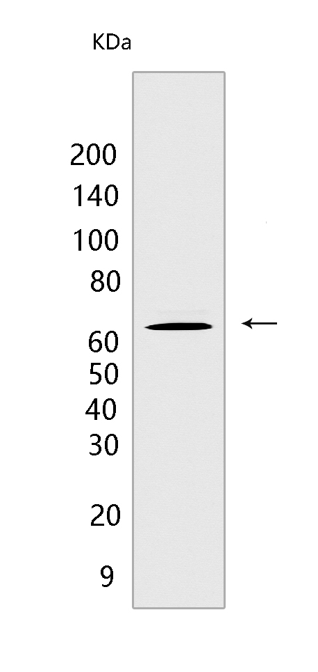
Western blot(SDS PAGE) analysis of extracts from Jurkat cells.Using LAG3 Rabbit mAb [M71Y] at dilution of 1:1000 incubated at 4℃ over night.
Product information
Protein names :LAG3,FDC,LAG3_HUMAN,Lymphocyte activation gene 3 protein [Cleaved into: Secreted lymphocyte activation gene 3 protein ]
UniProtID :P18627
MASS(da) :57,449
MW(kDa) :60-80 kDa
Form :Liquid
Purification :Protein A purification
Host :Rabbit
Isotype :IgG
sensitivity :Endogenous
Reactivity :Human
- ApplicationDilution
- 免疫印迹(WB)1:1000-2000
- The optimal dilutions should be determined by the end user
Specificity :Antibody is produced by immunizing animals with a synthetic peptide at the sequence of Human LAG3
Storage :Antibody store in 10 mM PBS, 0.5mg/ml BSA, 50% glycerol. Shipped at 4°C. Store at-20°C or -80°C. Products are valid for one natural year of receipt.Avoid repeated freeze / thaw cycles.
WB Positive detected :Jurkat cells
Function : Lymphocyte activation gene 3 protein: Inhibitory receptor on antigen activated T-cells (PubMed:7805750, PubMed:8647185, PubMed:20421648). Delivers inhibitory signals upon binding to ligands, such as FGL1 (By similarity). FGL1 constitutes a major ligand of LAG3 and is responsible for LAG3 T-cell inhibitory function (By similarity). Following TCR engagement, LAG3 associates with CD3-TCR in the immunological synapse and directly inhibits T-cell activation (By similarity). May inhibit antigen-specific T-cell activation in synergy with PDCD1/PD-1, possibly by acting as a coreceptor for PDCD1/PD-1 (By similarity). Negatively regulates the proliferation, activation, effector function and homeostasis of both CD8(+) and CD4(+) T-cells (PubMed:7805750, PubMed:8647185, PubMed:20421648). Also mediates immune tolerance: constitutively expressed on a subset of regulatory T-cells (Tregs) and contributes to their suppressive function (By similarity). Also acts as a negative regulator of plasmacytoid dendritic cell (pDCs) activation (By similarity). Binds MHC class II (MHC-II),the precise role of MHC-II-binding is however unclear (PubMed:8647185).., [Secreted lymphocyte activation gene 3 protein]: May function as a ligand for MHC class II (MHC-II) on antigen-presenting cells (APC), promoting APC activation/maturation and driving Th1 immune response..
Tissue specificity :Primarily expressed in activated T-cells and a subset of natural killer (NK) cells..
Subcellular locationi :[Lymphocyte activation gene 3 protein]: Cell membrane,Single-pass type I membrane protein.,[Secreted lymphocyte activation gene 3 protein]: Secreted.
IMPORTANT: For western blots, incubate membrane with diluted primary antibody in 1% w/v BSA, 1X TBST at 4°C overnight.


