GOLPH2 Rabbit mAb [RZ2Y]Cat NO.: A16562
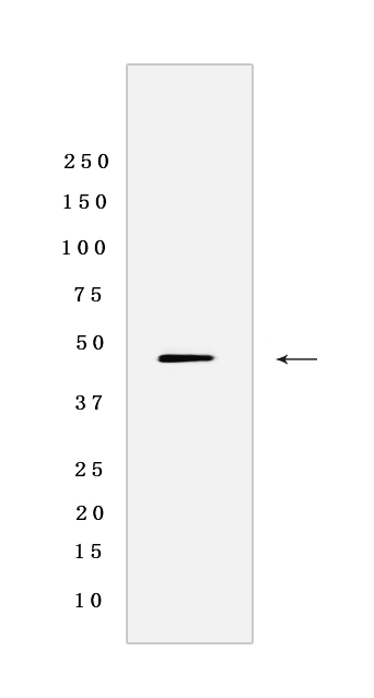
Western blot analysis of extracts from Huh7 cells lyastes.using GOLPH2 Rabbit mAb [RZ2Y] at dilution of 1:1000 incubated at 4℃ over night
Product information
Protein names :GOLM1,C9orf155,GOLPH2,PSEC0242,UNQ686/PRO1326,GOLM1_HUMAN,Golgi membrane protein 1
UniProtID :Q8NBJ4
MASS(da) :45,333
MW(kDa) :45 kDa
Form :Liquid
Purification :Protein A purification
Host :Rabbit
Isotype :IgG
sensitivity :Endogenous
Reactivity :Human
- ApplicationDilution
- 免疫印迹(WB)1:1000-2000
- 免疫组化(IHC)1:100,
- The optimal dilutions should be determined by the end user
Specificity :Antibody is produced by immunizing animals with a synthetic peptide of Human GOLPH2.
Storage :Antibody store in 10 mM PBS, 0.5mg/ml BSA, 50% glycerol. Shipped at 4°C. Store at-20°C or -80°C. Products are valid for one natural year of receipt.Avoid repeated freeze / thaw cycles.
WB Positive detected :Huh7 cells lyastes
Function : Unknown. Cellular response protein to viral infection.
Tissue specificity :Widely expressed. Highly expressed in colon, prostate, trachea and stomach. Expressed at lower level in testis, muscle, lymphoid tissues, white blood cells and spleen. Predominantly expressed by cells of the epithelial lineage. Expressed at low level in normal liver. Expression significantly increases in virus (HBV, HCV) infected liver. Expression does not increase in liver disease due to non-viral causes (alcohol-induced liver disease, autoimmune hepatitis). Increased expression in hepatocytes appears to be a general feature of advanced liver disease. In liver tissue from patients with adult giant-cell hepatitis (GCH), it is strongly expressed in hepatocytes-derived syncytial giant cells. Constitutively expressed by biliary epithelial cells but not by hepatocytes..
Subcellular locationi :Golgi apparatus, cis-Golgi network membrane,Single-pass type II membrane protein.
IMPORTANT: For western blots, incubate membrane with diluted primary antibody in 1% w/v BSA, 1X TBST at 4°C overnight.


