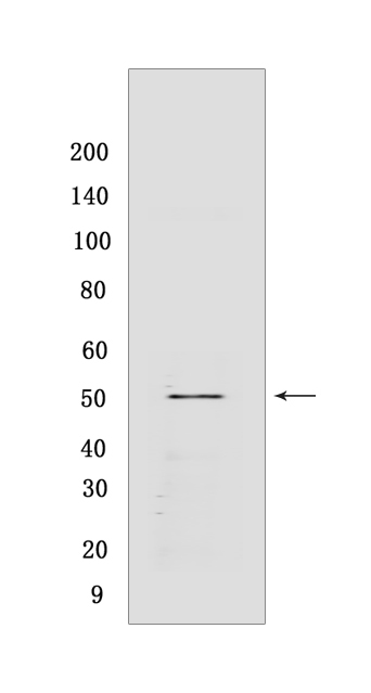
LAIR-1 Rabbit mAb [G9GL]Cat NO.: A87334
Western blot(SDS PAGE) analysis of extracts from THP-1 cells.Using LAIR-1 Rabbit mAb [G9GL] at dilution of 1:1000 incubated at 4℃ over night.
Product information
Protein names :LAIR1,CD305,LAIR1_HUMAN,Leukocyte-associated immunoglobulin-like receptor 1
UniProtID :Q6GTX8
MASS(da) :31,412
MW(kDa) :50 kDa
Form :Liquid
Purification :Protein A purification
Host :Rabbit
Isotype :IgG
sensitivity :Endogenous
Reactivity :Human
- ApplicationDilution
- 免疫印迹(WB)1:1000-2000
- The optimal dilutions should be determined by the end user
Specificity :Antibody is produced by immunizing animals with a synthetic peptide at the sequence of Human LAIR-1
Storage :Antibody store in 10 mM PBS, 0.5mg/ml BSA, 50% glycerol. Shipped at 4°C. Store at-20°C or -80°C. Products are valid for one natural year of receipt.Avoid repeated freeze / thaw cycles.
WB Positive detected :THP-1 cells
Function : Functions as an inhibitory receptor that plays a constitutive negative regulatory role on cytolytic function of natural killer (NK) cells, B-cells and T-cells. Activation by Tyr phosphorylation results in recruitment and activation of the phosphatases PTPN6 and PTPN11. It also reduces the increase of intracellular calcium evoked by B-cell receptor ligation. May also play its inhibitory role independently of SH2-containing phosphatases. Modulates cytokine production in CD4+ T-cells, down-regulating IL2 and IFNG production while inducing secretion of transforming growth factor beta. Down-regulates also IgG and IgE production in B-cells as well as IL8, IL10 and TNF secretion. Inhibits proliferation and induces apoptosis in myeloid leukemia cell lines as well as prevents nuclear translocation of NF-kappa-B p65 subunit/RELA and phosphorylation of I-kappa-B alpha/CHUK in these cells. Inhibits the differentiation of peripheral blood precursors towards dendritic cells..
Tissue specificity :Expressed on the majority of peripheral mononuclear cells, including natural killer (NK) cells, T-cells, B-cells, monocytes, and dendritic cells. Highly expressed in naive T-cells and B-cells but no expression on germinal center B-cells. Abnormally low expression in naive B-cells from HIV-1 infected patients. Very low expression in NK cells from a patient with chronic active Epstein-Barr virus infection..
Subcellular locationi :Cell membrane,Single-pass type I membrane protein.
IMPORTANT: For western blots, incubate membrane with diluted primary antibody in 1% w/v BSA, 1X TBST at 4°C overnight.


