
Dysbindin Rabbit mAb [HM25]Cat NO.: A54702
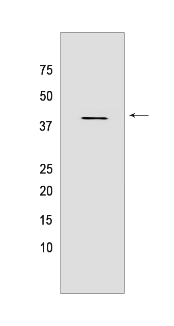
Western blot analysis of extracts from 293T cells lyastes.using Dysbindin Rabbit mAb [HM25] at dilution of 1:1000 incubated at 4℃ over night
Product information
Protein names :DTNBP1,BLOC1S8,My031,DTBP1_HUMAN,Dysbindin
UniProtID :Q96EV8
MASS(da) :39,493
MW(kDa) :39 kDa
Form :Liquid
Purification :Protein A purification
Host :Rabbit
Isotype :IgG
sensitivity :Endogenous
Reactivity :Human
- ApplicationDilution
- 免疫印迹(WB)1:1000-2000
- 免疫组化(IHC)1:100,
- The optimal dilutions should be determined by the end user
Specificity :Antibody is produced by immunizing animals with a synthetic peptide of Human Dysbindin.
Storage :Antibody store in 10 mM PBS, 0.5mg/ml BSA, 50% glycerol. Shipped at 4°C. Store at-20°C or -80°C. Products are valid for one natural year of receipt.Avoid repeated freeze / thaw cycles.
WB Positive detected :293T cells lyastes
Function : Component of the BLOC-1 complex, a complex that is required for normal biogenesis of lysosome-related organelles (LRO), such as platelet dense granules and melanosomes. In concert with the AP-3 complex, the BLOC-1 complex is required to target membrane protein cargos into vesicles assembled at cell bodies for delivery into neurites and nerve terminals. The BLOC-1 complex, in association with SNARE proteins, is also proposed to be involved in neurite extension. Associates with the BLOC-2 complex to facilitate the transport of TYRP1 independent of AP-3 function. Plays a role in synaptic vesicle trafficking and in neurotransmitter release. Plays a role in the regulation of cell surface exposure of DRD2. May play a role in actin cytoskeleton reorganization and neurite outgrowth. May modulate MAPK8 phosphorylation. Appears to promote neuronal transmission and viability through regulating the expression of SNAP25 and SYN1, modulating PI3-kinase-Akt signaling and influencing glutamatergic release. Regulates the expression of SYN1 through binding to its promoter. Modulates prefrontal cortical activity via the dopamine/D2 pathway..
Tissue specificity :Detected in brain, in neurons and in neuropil. Isoform 1 is expressed in the cerebral cortex, and hippocampal frontal (HF). Specific expression in the posterior half of the superior temporal gyrus (pSTG). Higher expression of isoform 2 and 3 in the HF than in the pSTG while isoform 1 shows no difference in expression in these areas. In the HF, detected in dentate gyrus (DG) and in pyramidal cells of hippocampus CA2 and CA3 (at protein level). Expressed in all principal neuronal populations of the HF, namely pyramidal neurons in the subiculum and CA1-3, granule cells in the dense cell layer of the DG (DGg), and polymorph cells in the hilus of the DG (DGh). Maximal levels in CA2, CA3, and DGh. Isoform 2 not expressed in the cerebral cortex..
Subcellular locationi :[Isoform 1]: Cytoplasm. Cytoplasmic vesicle membrane,Peripheral membrane protein,Cytoplasmic side. Endosome membrane,Peripheral membrane protein,Cytoplasmic side. Melanosome membrane,Peripheral membrane protein,Cytoplasmic side. Cell junction, synapse, postsynaptic density. Endoplasmic reticulum. Nucleus.
IMPORTANT: For western blots, incubate membrane with diluted primary antibody in 1% w/v BSA, 1X TBST at 4°C overnight.
-
About
-
Product
-
Law
-
Literature
-

WeChat scan code follow us