
ARG1 Rabbit mAb [j3zA]Cat NO.: A98832
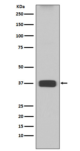
Western blot(SDS PAGE) analysis of extracts from Mouse liver lysate.Using ARG1 Rabbit mAb [j3zA]at dilution of 1:1000 incubated at 4℃ over night.
Product information
Protein names :ARG1; Arginase-1; Liver-type arginase; Type I arginase; Arginase liver; Liver Arginase;
UniProtID :P05089
MASS(da) :34,735
MW(kDa) :35kDa
Form :Liquid
Purification :Affinity-chromatography
Host :Rabbit
Isotype : IgG
sensitivity :Endogenous
Reactivity :Human,Mouse,Rat
- ApplicationDilution
- 免疫印迹(WB)1:1000-2000
- The optimal dilutions should be determined by the end user
Specificity :Antibody is produced by immunizing animals with A synthesized peptide derived from human ARG1
Storage :Antibody store in 10 mM PBS, 0.5mg/ml BSA, 50% glycerol. Shipped at 4°C. Store at-20°C or -80°C. Products are valid for one natural year of receipt.Avoid repeated freeze / thaw cycles.
WB Positive detected :Mouse liver lysate.
Function : Key element of the urea cycle converting L-arginine to urea and L-ornithine, which is further metabolized into metabolites proline and polyamides that drive collagen synthesis and bioenergetic pathways critical for cell proliferation, respectively,the urea cycle takes place primarily in the liver and, to a lesser extent, in the kidneys.., Functions in L-arginine homeostasis in nonhepatic tissues characterized by the competition between nitric oxide synthase (NOS) and arginase for the available intracellular substrate arginine. Arginine metabolism is a critical regulator of innate and adaptive immune responses. Involved in an antimicrobial effector pathway in polymorphonuclear granulocytes (PMN). Upon PMN cell death is liberated from the phagolysosome and depletes arginine in the microenvironment leading to suppressed T cell and natural killer (NK) cell proliferation and cytokine secretion (PubMed:15546957, PubMed:16709924, PubMed:19380772). In group 2 innate lymphoid cells (ILC2s) promotes acute type 2 inflammation in the lung and is involved in optimal ILC2 proliferation but not survival (By similarity). In humans, the immunological role in the monocytic/macrophage/dendritic cell (DC) lineage is unsure..
Tissue specificity :Within the immune system initially reported to be selectively expressed in granulocytes (polymorphonuclear leukocytes [PMNs]) (PubMed:15546957). Also detected in macrophages mycobacterial granulomas (PubMed:23749634). Expressed in group2 innate lymphoid cells (ILC2s) during lung disease (PubMed:27043409)..
Subcellular locationi :Cytoplasm. Cytoplasmic granule.
IMPORTANT: For western blots, incubate membrane with diluted primary antibody in 1% w/v BSA, 1X TBST at 4°C overnight.
-
About
-
Product
-
Law
-
Literature
-

WeChat scan code follow us