
TLR2 Rabbit mAb [5rt7]Cat NO.: A58502
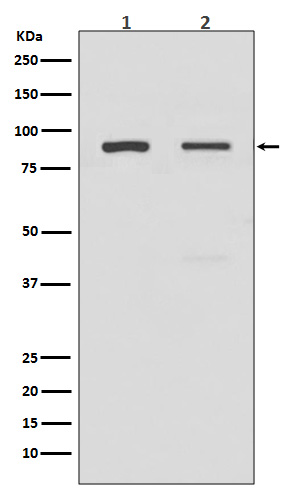
Western blot(SDS PAGE) analysis of extracts from (1) A549 cell lysate; (2) HeLa cell lysate.Using TLR2 Rabbit mAb [5rt7]at dilution of 1:1000 incubated at 4℃ over night.
Product information
Protein names :TLR2;CD282;TIL4;Toll-like receptor 2;Toll/interleukin-1 receptor-like protein 4;
UniProtID :O60603
MASS(da) :89,838
MW(kDa) :90kDa
Form :Liquid
Purification :Affinity-chromatography
Host :Rabbit
Isotype : IgG
sensitivity :Endogenous
Reactivity :Human,Mouse,Rat
- ApplicationDilution
- 免疫印迹(WB)1:1000-2000
- The optimal dilutions should be determined by the end user
Specificity :Antibody is produced by immunizing animals with A synthesized peptide derived from human TLR2
Storage :Antibody store in 10 mM PBS, 0.5mg/ml BSA, 50% glycerol. Shipped at 4°C. Store at-20°C or -80°C. Products are valid for one natural year of receipt.Avoid repeated freeze / thaw cycles.
WB Positive detected :(1) A549 cell lysate; (2) HeLa cell lysate.
Function : Cooperates with LY96 to mediate the innate immune response to bacterial lipoproteins and other microbial cell wall components. Cooperates with TLR1 or TLR6 to mediate the innate immune response to bacterial lipoproteins or lipopeptides (PubMed:21078852, PubMed:17889651). Acts via MYD88 and TRAF6, leading to NF-kappa-B activation, cytokine secretion and the inflammatory response. May also activate immune cells and promote apoptosis in response to the lipid moiety of lipoproteins (PubMed:10426995, PubMed:10426996). Recognizes mycoplasmal macrophage-activating lipopeptide-2kD (MALP-2), soluble tuberculosis factor (STF), phenol-soluble modulin (PSM) and B.burgdorferi outer surface protein A lipoprotein (OspA-L) cooperatively with TLR6 (PubMed:11441107). Stimulation of monocytes in vitro with M.tuberculosis PstS1 induces p38 MAPK and ERK1/2 activation primarily via this receptor, but also partially via TLR4 (PubMed:16622205). MAPK activation in response to bacterial peptidoglycan also occurs via this receptor (PubMed:16622205). Acts as a receptor for M.tuberculosis lipoproteins LprA, LprG, LpqH and PstS1, some lipoproteins are dependent on other coreceptors (TLR1, CD14 and/or CD36),the lipoproteins act as agonists to modulate antigen presenting cell functions in response to the pathogen (PubMed:19362712). M.tuberculosis HSP70 (dnaK) but not HSP65 (groEL-2) acts via this protein to stimulate NF-kappa-B expression (PubMed:15809303). Recognizes M.tuberculosis major T-antigen EsxA (ESAT-6) which inhibits downstream MYD88-dependent signaling (shown in mouse) (By similarity). Forms activation clusters composed of several receptors depending on the ligand, these clusters trigger signaling from the cell surface and subsequently are targeted to the Golgi in a lipid-raft dependent pathway. Forms the cluster TLR2:TLR6:CD14:CD36 in response to diacylated lipopeptides and TLR2:TLR1:CD14 in response to triacylated lipopeptides (PubMed:16880211). Required for normal uptake of M.tuberculosis, a process that is inhibited by M.tuberculosis LppM (By similarity)..
Tissue specificity :Highly expressed in peripheral blood leukocytes, in particular in monocytes, in bone marrow, lymph node and in spleen. Also detected in lung and in fetal liver. Levels are low in other tissues.
Subcellular locationi :Membrane,Single-pass type I membrane protein. Cytoplasmic vesicle, phagosome membrane,Single-pass type I membrane protein. Membrane raft.
IMPORTANT: For western blots, incubate membrane with diluted primary antibody in 1% w/v BSA, 1X TBST at 4°C overnight.
-
About
-
Product
-
Law
-
Literature
-

WeChat scan code follow us