
P-Smad3 (S423 + S425) Rabbit mAb [g4Bi]Cat NO.: A64266
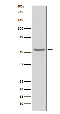
Western blot(SDS PAGE) analysis of extracts from A549 cell lysate treated with TGF-ß1.Using P-Smad3 (S423 + S425) Rabbit mAb [g4Bi]at dilution of 1:1000 incubated at 4℃ over night.
Product information
Protein names :JV15-2, MAD-3, MADH3, Mad3, Mothers against DPP homolog 3, Mothers against decapentaplegic homolog 3, SMAD 3, Smad 3
UniProtID :P84022
MASS(da) :48,081
MW(kDa) :55kDa
Form :Liquid
Purification :Affinity-chromatography
Host :Rabbit
Isotype : IgG
sensitivity :Endogenous
Reactivity :Human Mouse
- ApplicationDilution
- 免疫印迹(WB)1:1000-2000
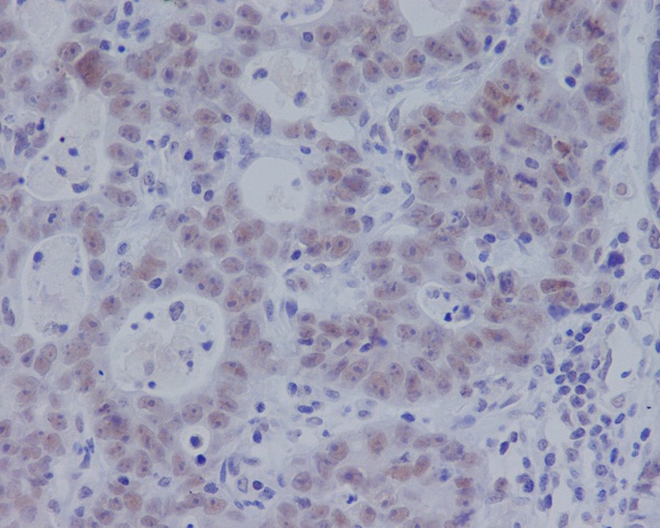
- 免疫组化(IHC)1:100
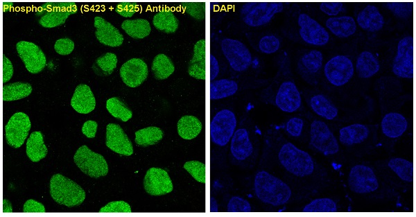
- 免疫荧光(ICC/IF)1:100
- The optimal dilutions should be determined by the end user
Specificity :Antibody is produced by immunizing animals with A synthesized peptide derived from human Phospho-Smad3 (S423 + S425)
Storage :Antibody store in 10 mM PBS, 0.5mg/ml BSA, 50% glycerol. Shipped at 4°C. Store at-20°C or -80°C. Products are valid for one natural year of receipt.Avoid repeated freeze / thaw cycles.
WB Positive detected :A549 cell lysate treated with TGF-ß1.
Function : Receptor-regulated SMAD (R-SMAD) that is an intracellular signal transducer and transcriptional modulator activated by TGF-beta (transforming growth factor) and activin type 1 receptor kinases. Binds the TRE element in the promoter region of many genes that are regulated by TGF-beta and, on formation of the SMAD3/SMAD4 complex, activates transcription. Also can form a SMAD3/SMAD4/JUN/FOS complex at the AP-1/SMAD site to regulate TGF-beta-mediated transcription. Has an inhibitory effect on wound healing probably by modulating both growth and migration of primary keratinocytes and by altering the TGF-mediated chemotaxis of monocytes. This effect on wound healing appears to be hormone-sensitive. Regulator of chondrogenesis and osteogenesis and inhibits early healing of bone fractures. Positively regulates PDPK1 kinase activity by stimulating its dissociation from the 14-3-3 protein YWHAQ which acts as a negative regulator..
Subcellular locationi :Cytoplasm. Nucleus.
IMPORTANT: For western blots, incubate membrane with diluted primary antibody in 1% w/v BSA, 1X TBST at 4°C overnight.
-
About
-
Product
-
Law
-
Literature
-

WeChat scan code follow us