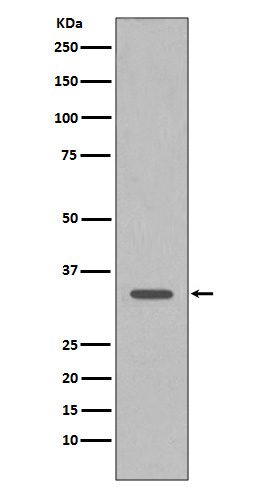
CDK1 Rabbit mAb [4WuL]Cat NO.: A18280
Western blot(SDS PAGE) analysis of extracts from HeLa cell lysate.Using CDK1 Rabbit mAb [4WuL]at dilution of 1:1000 incubated at 4℃ over night.
Product information
Protein names :CDC2; CDC28A; P34CDC2; CDK1;
UniProtID :P06493
MASS(da) :34,095
MW(kDa) :34kDa
Form :Liquid
Purification :Affinity-chromatography
Host :Rabbit
Isotype : IgG
sensitivity :Endogenous
Reactivity :Human,Mouse,Rat
- ApplicationDilution
- 免疫印迹(WB)1:1000-2000
- The optimal dilutions should be determined by the end user
Specificity :Antibody is produced by immunizing animals with A synthesized peptide derived from human CDK1
Storage :Antibody store in 10 mM PBS, 0.5mg/ml BSA, 50% glycerol. Shipped at 4°C. Store at-20°C or -80°C. Products are valid for one natural year of receipt.Avoid repeated freeze / thaw cycles.
WB Positive detected :HeLa cell lysate.
Function : Plays a key role in the control of the eukaryotic cell cycle by modulating the centrosome cycle as well as mitotic onset,promotes G2-M transition, and regulates G1 progress and G1-S transition via association with multiple interphase cyclins (PubMed:16407259, PubMed:17459720, PubMed:16933150, PubMed:18356527, PubMed:19509060, PubMed:20171170, PubMed:19917720, PubMed:20937773, PubMed:20935635, PubMed:21063390, PubMed:23355470, PubMed:23601106, PubMed:23602554, PubMed:25556658, PubMed:26829474, PubMed:30704899). Required in higher cells for entry into S-phase and mitosis (PubMed:16407259, PubMed:17459720, PubMed:16933150, PubMed:18356527, PubMed:19509060, PubMed:20171170, PubMed:19917720, PubMed:20937773, PubMed:20935635, PubMed:21063390, PubMed:23355470, PubMed:23601106, PubMed:23602554, PubMed:25556658). Phosphorylates PARVA/actopaxin, APC, AMPH, APC, BARD1, Bcl-xL/BCL2L1, BRCA2, CALD1, CASP8, CDC7, CDC20, CDC25A, CDC25C, CC2D1A, CENPA, CSNK2 proteins/CKII, FZR1/CDH1, CDK7, CEBPB, CHAMP1, DMD/dystrophin, EEF1 proteins/EF-1, EZH2, KIF11/EG5, EGFR, FANCG, FOS, GFAP, GOLGA2/GM130, GRASP1, UBE2A/hHR6A, HIST1H1 proteins/histone H1, HMGA1, HIVEP3/KRC, KAT5, LMNA, LMNB, LMNC, LBR, LATS1, MAP1B, MAP4, MARCKS, MCM2, MCM4, MKLP1, MYB, NEFH, NFIC, NPC/nuclear pore complex, PITPNM1/NIR2, NPM1, NCL, NUCKS1, NPM1/numatrin, ORC1, PRKAR2A, EEF1E1/p18, EIF3F/p47, p53/TP53, NONO/p54NRB, PAPOLA, PLEC/plectin, RB1, TPPP, UL40/R2, RAB4A, RAP1GAP, RCC1, RPS6KB1/S6K1, KHDRBS1/SAM68, ESPL1, SKI, BIRC5/survivin, STIP1, TEX14, beta-tubulins, MAPT/TAU, NEDD1, VIM/vimentin, TK1, FOXO1, RUNX1/AML1, SAMHD1, SIRT2, CGAS and RUNX2 (PubMed:16407259, PubMed:17459720, PubMed:16933150, PubMed:18356527, PubMed:19509060, PubMed:20171170, PubMed:19917720, PubMed:20937773, PubMed:20935635, PubMed:21063390, PubMed:23355470, PubMed:23601106, PubMed:23602554, PubMed:25556658, PubMed:32351706, PubMed:26829474, PubMed:30704899). CDK1/CDC2-cyclin-B controls pronuclear union in interphase fertilized eggs (PubMed:18480403, PubMed:20360007). Essential for early stages of embryonic development (PubMed:18480403, PubMed:20360007). During G2 and early mitosis, CDC25A/B/C-mediated dephosphorylation activates CDK1/cyclin complexes which phosphorylate several substrates that trigger at least centrosome separation, Golgi dynamics, nuclear envelope breakdown and chromosome condensation (PubMed:18480403, PubMed:20360007). Once chromosomes are condensed and aligned at the metaphase plate, CDK1 activity is switched off by WEE1- and PKMYT1-mediated phosphorylation to allow sister chromatid separation, chromosome decondensation, reformation of the nuclear envelope and cytokinesis (PubMed:18480403, PubMed:20360007). Inactivated by PKR/EIF2AK2- and WEE1-mediated phosphorylation upon DNA damage to stop cell cycle and genome replication at the G2 checkpoint thus facilitating DNA repair (PubMed:20360007). Reactivated after successful DNA repair through WIP1-dependent signaling leading to CDC25A/B/C-mediated dephosphorylation and restoring cell cycle progression (PubMed:20395957). In proliferating cells, CDK1-mediated FOXO1 phosphorylation at the G2-M phase represses FOXO1 interaction with 14-3-3 proteins and thereby promotes FOXO1 nuclear accumulation and transcription factor activity, leading to cell death of postmitotic neurons (PubMed:18356527). The phosphorylation of beta-tubulins regulates microtubule dynamics during mitosis (PubMed:16371510). NEDD1 phosphorylation promotes PLK1-mediated NEDD1 phosphorylation and subsequent targeting of the gamma-tubulin ring complex (gTuRC) to the centrosome, an important step for spindle formation (PubMed:19509060). In addition, CC2D1A phosphorylation regulates CC2D1A spindle pole localization and association with SCC1/RAD21 and centriole cohesion during mitosis (PubMed:20171170). The phosphorylation of Bcl-xL/BCL2L1 after prolongated G2 arrest upon DNA damage triggers apoptosis (PubMed:19917720). In contrast, CASP8 phosphorylation during mitosis prevents its activation by proteolysis and subsequent apoptosis (PubMed:20937773). This phosphorylation occurs in cancer cell lines, as well as in primary breast tissues and lymphocytes (PubMed:20937773). EZH2 phosphorylation promotes H3K27me3 maintenance and epigenetic gene silencing (PubMed:20935635). CALD1 phosphorylation promotes Schwann cell migration during peripheral nerve regeneration (By similarity). CDK1-cyclin-B complex phosphorylates NCKAP5L and mediates its dissociation from centrosomes during mitosis (PubMed:26549230). Regulates the amplitude of the cyclic expression of the core clock gene ARNTL/BMAL1 by phosphorylating its transcriptional repressor NR1D1, and this phosphorylation is necessary for SCF(FBXW7)-mediated ubiquitination and proteasomal degradation of NR1D1 (PubMed:27238018). Phosphorylates EML3 at 'Thr-881' which is essential for its interaction with HAUS augmin-like complex and TUBG1 (PubMed:30723163). Phosphorylates CGAS during mitosis, leading to its inhibition, thereby preventing CGAS activation by self DNA during mitosis (PubMed:32351706).., (Microbial infection) Acts as a receptor for hepatitis C virus (HCV) in hepatocytes and facilitates its cell entry..
Tissue specificity :Isoform 2 is found in breast cancer tissues.
Subcellular locationi :Nucleus. Cytoplasm. Mitochondrion. Cytoplasm, cytoskeleton, microtubule organizing center, centrosome. Cytoplasm, cytoskeleton, spindle.
IMPORTANT: For western blots, incubate membrane with diluted primary antibody in 1% w/v BSA, 1X TBST at 4°C overnight.


