
pan Cadherin Rabbit mAb [MDu1]Cat NO.: A18062
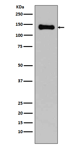
Western blot(SDS PAGE) analysis of extracts from C6 cell lysate.Using pan Cadherin Rabbit mAb [MDu1]at dilution of 1:1000 incubated at 4℃ over night.
Product information
Protein names :Cadherin;CDH3;CDHP;Cadherin-1;Cadherin-2;Cadherin-3;Cadherin-4;Cadherin-5;CAM 120/80;CD144;CD324;CD325;Cdh4;CDH5;CDHE;CDHN;E-cadherin;N-cadherin;NCAD;Neural cadherin;P-cadherin;Uvomorulin;Vascular endothelial cadherin
UniProtID :P12830/P19022/P22223/P33151/P55283/P39038
MASS(da) :97,456
MW(kDa) :140kDa
Form :Liquid
Purification :Affinity-chromatography
Host :Rabbit
Isotype : IgG
sensitivity :Endogenous
Reactivity :Human,Mouse,Rat
- ApplicationDilution
- 免疫印迹(WB)1:1000-2000
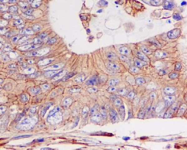
- 免疫组化(IHC)1:100
- 免疫荧光(ICC/IF)1:100
- The optimal dilutions should be determined by the end user
Specificity :Antibody is produced by immunizing animals with A synthesized peptide derived from human pan Cadherin
Storage :Antibody store in 10 mM PBS, 0.5mg/ml BSA, 50% glycerol. Shipped at 4°C. Store at-20°C or -80°C. Products are valid for one natural year of receipt.Avoid repeated freeze / thaw cycles.
WB Positive detected :C6 cell lysate.
Function : Cadherins are calcium-dependent cell adhesion proteins (PubMed:11976333). They preferentially interact with themselves in a homophilic manner in connecting cells,cadherins may thus contribute to the sorting of heterogeneous cell types. CDH1 is involved in mechanisms regulating cell-cell adhesions, mobility and proliferation of epithelial cells (PubMed:11976333). Has a potent invasive suppressor role. It is a ligand for integrin alpha-E/beta-7.., E-Cad/CTF2 promotes non-amyloidogenic degradation of Abeta precursors. Has a strong inhibitory effect on APP C99 and C83 production.., (Microbial infection) Serves as a receptor for Listeria monocytogenes,internalin A (InlA) binds to this protein and promotes uptake of the bacteria..
Tissue specificity :Non-neural epithelial tissues.
Subcellular locationi :Cell junction, adherens junction. Cell membrane,Single-pass type I membrane protein. Endosome. Golgi apparatus, trans-Golgi network.
IMPORTANT: For western blots, incubate membrane with diluted primary antibody in 1% w/v BSA, 1X TBST at 4°C overnight.
-
About
-
Product
-
Law
-
Literature
-

WeChat scan code follow us