
Caldesmon Rabbit mAb [fMC2]Cat NO.: A95930
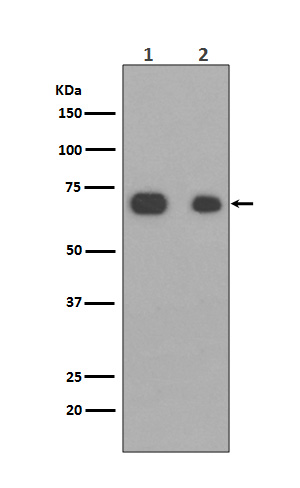
Western blot(SDS PAGE) analysis of extracts from (1)NIH3T3 cell lysate; (2)HeLa cell lysate.Using Caldesmon Rabbit mAb [fMC2]at dilution of 1:1000 incubated at 4℃ over night.
Product information
Protein names :CAD; CALD1; CDM; L-caldesmon; Non-muscle caldesmon;
UniProtID :Q05682
MASS(da) :93,231
MW(kDa) :70kDa
Form :Liquid
Purification :Affinity-chromatography
Host :Rabbit
Isotype : IgG
sensitivity :Endogenous
Reactivity :Human,Mouse,Rat
- ApplicationDilution
- 免疫印迹(WB)1:1000-2000
- 免疫组化(IHC)1:100
- 免疫荧光(ICC/IF)1:100
- The optimal dilutions should be determined by the end user
Specificity :Antibody is produced by immunizing animals with A synthesized peptide derived from human Caldesmon
Storage :Antibody store in 10 mM PBS, 0.5mg/ml BSA, 50% glycerol. Shipped at 4°C. Store at-20°C or -80°C. Products are valid for one natural year of receipt.Avoid repeated freeze / thaw cycles.
WB Positive detected :(1)NIH3T3 cell lysate; (2)HeLa cell lysate.
Function : Actin- and myosin-binding protein implicated in the regulation of actomyosin interactions in smooth muscle and nonmuscle cells (could act as a bridge between myosin and actin filaments). Stimulates actin binding of tropomyosin which increases the stabilization of actin filament structure. In muscle tissues, inhibits the actomyosin ATPase by binding to F-actin. This inhibition is attenuated by calcium-calmodulin and is potentiated by tropomyosin. Interacts with actin, myosin, two molecules of tropomyosin and with calmodulin. Also plays an essential role during cellular mitosis and receptor capping. Involved in Schwann cell migration during peripheral nerve regeneration (By similarity)..
Tissue specificity :High-molecular-weight caldesmon (isoform 1) is predominantly expressed in smooth muscles, whereas low-molecular-weight caldesmon (isoforms 2, 3, 4 and 5) are widely distributed in non-muscle tissues and cells. Not expressed in skeletal muscle or heart.
Subcellular locationi :Cytoplasm, cytoskeleton. Cytoplasm, myofibril. Cytoplasm, cytoskeleton, stress fiber.
IMPORTANT: For western blots, incubate membrane with diluted primary antibody in 1% w/v BSA, 1X TBST at 4°C overnight.
-
About
-
Product
-
Law
-
Literature
-

WeChat scan code follow us