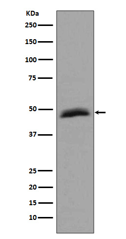
Aurora A Rabbit mAb [8Hkv]Cat NO.: A51437
Western blot(SDS PAGE) analysis of extracts from HepG2 cell lysate.Using Aurora A Rabbit mAb [8Hkv]at dilution of 1:1000 incubated at 4℃ over night.
Product information
Protein names :AIK, ARK1, AYK1, Aurora-A, Aurora-related kinase 1, BTAK,IAK1, Ipl1- and aurora-related kinase 1, STK15, STK6, Serine/threonine kinase 15
UniProtID :O14965
MASS(da) :45,823
MW(kDa) :46kDa
Form :Liquid
Purification :Affinity-chromatography
Host :Rabbit
Isotype : IgG
sensitivity :Endogenous
Reactivity :Human Mouse
- ApplicationDilution
- 免疫印迹(WB)1:1000-2000
- 免疫荧光(ICC/IF)1:100
- The optimal dilutions should be determined by the end user
Specificity :Antibody is produced by immunizing animals with A synthesized peptide derived from human Aurora A
Storage :Antibody store in 10 mM PBS, 0.5mg/ml BSA, 50% glycerol. Shipped at 4°C. Store at-20°C or -80°C. Products are valid for one natural year of receipt.Avoid repeated freeze / thaw cycles.
WB Positive detected :HepG2 cell lysate.
Function : Mitotic serine/threonine kinase that contributes to the regulation of cell cycle progression (PubMed:26246606, PubMed:12390251, PubMed:18615013, PubMed:11039908, PubMed:17125279, PubMed:17360485). Associates with the centrosome and the spindle microtubules during mitosis and plays a critical role in various mitotic events including the establishment of mitotic spindle, centrosome duplication, centrosome separation as well as maturation, chromosomal alignment, spindle assembly checkpoint, and cytokinesis (PubMed:26246606, PubMed:14523000). Required for normal spindle positioning during mitosis and for the localization of NUMA1 and DCTN1 to the cell cortex during metaphase (PubMed:27335426). Required for initial activation of CDK1 at centrosomes (PubMed:13678582, PubMed:15128871). Phosphorylates numerous target proteins, including ARHGEF2, BORA, BRCA1, CDC25B, DLGP5, HDAC6, KIF2A, LATS2, NDEL1, PARD3, PPP1R2, PLK1, RASSF1, TACC3, p53/TP53 and TPX2 (PubMed:18056443, PubMed:15128871, PubMed:14702041, PubMed:11551964, PubMed:15147269, PubMed:15987997, PubMed:17604723, PubMed:18615013). Regulates KIF2A tubulin depolymerase activity (PubMed:19351716). Important for microtubule formation and/or stabilization (PubMed:18056443). Required for normal axon formation (PubMed:19812038). Plays a role in microtubule remodeling during neurite extension (PubMed:19668197). Also acts as a key regulatory component of the p53/TP53 pathway, and particularly the checkpoint-response pathways critical for oncogenic transformation of cells, by phosphorylating and destabilizing p53/TP53 (PubMed:14702041). Phosphorylates its own inhibitors, the protein phosphatase type 1 (PP1) isoforms, to inhibit their activity (PubMed:11551964). Necessary for proper cilia disassembly prior to mitosis (PubMed:17604723, PubMed:20643351). Regulates protein levels of the anti-apoptosis protein BIRC5 by suppressing the expression of the SCF(FBXL7) E3 ubiquitin-protein ligase substrate adapter FBXL7 through the phosphorylation of the transcription factor FOXP1 (PubMed:28218735)..
Tissue specificity :Highly expressed in testis and weakly in skeletal muscle, thymus and spleen. Also highly expressed in colon, ovarian, prostate, neuroblastoma, breast and cervical cancer cell lines.
Subcellular locationi :Cytoplasm, cytoskeleton, microtubule organizing center, centrosome. Cytoplasm, cytoskeleton, spindle pole. Cytoplasm, cytoskeleton, cilium basal body. Cytoplasm, cytoskeleton, microtubule organizing center, centrosome, centriole. Cell projection, neuron projection.
IMPORTANT: For western blots, incubate membrane with diluted primary antibody in 1% w/v BSA, 1X TBST at 4°C overnight.


