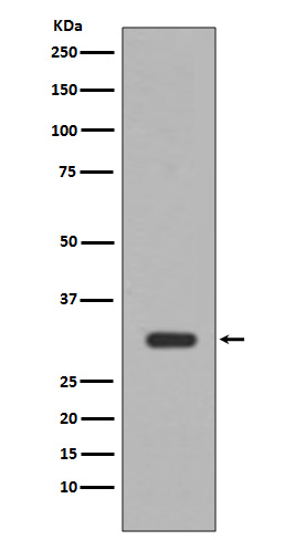
PIM1 Rabbit mAb [Lj93]Cat NO.: A78277
Western blot(SDS PAGE) analysis of extracts from Jurkat cell lysate.Using PIM1 Rabbit mAb [Lj93]at dilution of 1:1000 incubated at 4℃ over night.
Product information
Protein names :Oncogene PIM1; PIM; pim-1 kinase 44 kDa isoform; pim-1 oncogene; pim-1 oncogene (proviral integration site 1); PIM1;
UniProtID :P11309
MASS(da) :35,686
MW(kDa) :32kDa
Form :Liquid
Purification :Affinity-chromatography
Host :Rabbit
Isotype : IgG
sensitivity :Endogenous
Reactivity :Human Mouse
- ApplicationDilution
- 免疫印迹(WB)1:1000-2000
- 免疫组化(IHC)1:100
- 免疫荧光(ICC/IF)1:100
- The optimal dilutions should be determined by the end user
Specificity :Antibody is produced by immunizing animals with A synthesized peptide derived from human PIM1
Storage :Antibody store in 10 mM PBS, 0.5mg/ml BSA, 50% glycerol. Shipped at 4°C. Store at-20°C or -80°C. Products are valid for one natural year of receipt.Avoid repeated freeze / thaw cycles.
WB Positive detected :Jurkat cell lysate.
Function : Proto-oncogene with serine/threonine kinase activity involved in cell survival and cell proliferation and thus providing a selective advantage in tumorigenesis. Exerts its oncogenic activity through: the regulation of MYC transcriptional activity, the regulation of cell cycle progression and by phosphorylation and inhibition of proapoptotic proteins (BAD, MAP3K5, FOXO3). Phosphorylation of MYC leads to an increase of MYC protein stability and thereby an increase of transcriptional activity. The stabilization of MYC exerted by PIM1 might explain partly the strong synergism between these two oncogenes in tumorigenesis. Mediates survival signaling through phosphorylation of BAD, which induces release of the anti-apoptotic protein Bcl-X(L)/BCL2L1. Phosphorylation of MAP3K5, another proapoptotic protein, by PIM1, significantly decreases MAP3K5 kinase activity and inhibits MAP3K5-mediated phosphorylation of JNK and JNK/p38MAPK subsequently reducing caspase-3 activation and cell apoptosis. Stimulates cell cycle progression at the G1-S and G2-M transitions by phosphorylation of CDC25A and CDC25C. Phosphorylation of CDKN1A, a regulator of cell cycle progression at G1, results in the relocation of CDKN1A to the cytoplasm and enhanced CDKN1A protein stability. Promotes cell cycle progression and tumorigenesis by down-regulating expression of a regulator of cell cycle progression, CDKN1B, at both transcriptional and post-translational levels. Phosphorylation of CDKN1B, induces 14-3-3 proteins binding, nuclear export and proteasome-dependent degradation. May affect the structure or silencing of chromatin by phosphorylating HP1 gamma/CBX3. Acts also as a regulator of homing and migration of bone marrow cells involving functional interaction with the CXCL12-CXCR4 signaling axis. Also phosphorylates and activates the ATP-binding cassette transporter ABCG2, allowing resistance to drugs through their excretion from cells (PubMed:18056989). Promotes brown adipocyte differentiation (By similarity)..
Tissue specificity :Expressed primarily in cells of the hematopoietic and germline lineages. Isoform 1 and isoform 2 are both expressed in prostate cancer cell lines..
Subcellular locationi :[Isoform 1]: Cytoplasm. Nucleus.,[Isoform 2]: Cell membrane.
IMPORTANT: For western blots, incubate membrane with diluted primary antibody in 1% w/v BSA, 1X TBST at 4°C overnight.


