MLANA Rabbit mAb [92Xd]Cat NO.: A80443
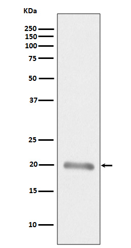
Western blot(SDS PAGE) analysis of extracts from Human melanoma tissue lysate.Using MLANA Rabbit mAb [92Xd]at dilution of 1:1000 incubated at 4℃ over night.
Product information
Protein names :Antigen LB39-AA; Antigen SK29-AA; MAR1; MART-1; MART1; melan-A; Melanoma antigen recognized by T-cells 1; MLANA; Protein Melan-A;
UniProtID :Q16655
MASS(da) :13,157
MW(kDa) :20kDa
Form :Liquid
Purification :Affinity-chromatography
Host :Rabbit
Isotype : IgG
sensitivity :Endogenous
Reactivity :Human
- ApplicationDilution
- 免疫印迹(WB)1:1000-2000
- 免疫组化(IHC)1:100
- 免疫荧光(ICC/IF)1:100
- The optimal dilutions should be determined by the end user
Specificity :Antibody is produced by immunizing animals with A synthesized peptide derived from human MLANA
Storage :Antibody store in 10 mM PBS, 0.5mg/ml BSA, 50% glycerol. Shipped at 4°C. Store at-20°C or -80°C. Products are valid for one natural year of receipt.Avoid repeated freeze / thaw cycles.
WB Positive detected :Human melanoma tissue lysate.
Function : Involved in melanosome biogenesis by ensuring the stability of GPR143. Plays a vital role in the expression, stability, trafficking, and processing of melanocyte protein PMEL, which is critical to the formation of stage II melanosomes..
Tissue specificity :Expression is restricted to melanoma and melanocyte cell lines and retina.
Subcellular locationi :Endoplasmic reticulum membrane,Single-pass type III membrane protein. Golgi apparatus. Golgi apparatus, trans-Golgi network membrane. Melanosome.
IMPORTANT: For western blots, incubate membrane with diluted primary antibody in 1% w/v BSA, 1X TBST at 4°C overnight.


