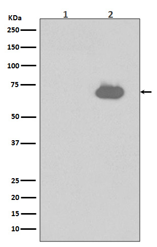
P-SHP2 (Y542) Rabbit mAb [jN6A]Cat NO.: A38301
Western blot(SDS PAGE) analysis of extracts from (1) NIH/3T3 cell lysates; (2) NIH/3T3 cell lysates treated with PDGF.Using P-SHP2 (Y542) Rabbit mAb [jN6A]at dilution of 1:1000 incubated at 4℃ over night.
Product information
Protein names :BPTP3; CFC; MGC14433; NS1; PTN11; PTP-1D; PTP-2C; PTP2C; PTPN11; SH-PTP2; SH-PTP3; SHP-2; Shp2; SHPTP2;
UniProtID :Q06124
MASS(da) :68,011
MW(kDa) :68kDa
Form :Liquid
Purification :Affinity-chromatography
Host :Rabbit
Isotype : IgG
sensitivity :Endogenous
Reactivity :Human Mouse
- ApplicationDilution
- 免疫印迹(WB)1:1000-2000
- 免疫荧光(ICC/IF)1:100
- The optimal dilutions should be determined by the end user
Specificity :Antibody is produced by immunizing animals with A synthesized peptide derived from human Phospho-SHP2 (Y542)
Storage :Antibody store in 10 mM PBS, 0.5mg/ml BSA, 50% glycerol. Shipped at 4°C. Store at-20°C or -80°C. Products are valid for one natural year of receipt.Avoid repeated freeze / thaw cycles.
WB Positive detected :(1) NIH/3T3 cell lysates; (2) NIH/3T3 cell lysates treated with PDGF.
Function : Acts downstream of various receptor and cytoplasmic protein tyrosine kinases to participate in the signal transduction from the cell surface to the nucleus (PubMed:10655584, PubMed:18559669, PubMed:18829466, PubMed:26742426, PubMed:28074573). Positively regulates MAPK signal transduction pathway (PubMed:28074573). Dephosphorylates GAB1, ARHGAP35 and EGFR (PubMed:28074573). Dephosphorylates ROCK2 at 'Tyr-722' resulting in stimulation of its RhoA binding activity (PubMed:18559669). Dephosphorylates CDC73 (PubMed:26742426). Dephosphorylates SOX9 on tyrosine residues, leading to inactivate SOX9 and promote ossification (By similarity)..
Tissue specificity :Widely expressed, with highest levels in heart, brain, and skeletal muscle..
Subcellular locationi :Cytoplasm. Nucleus.
IMPORTANT: For western blots, incubate membrane with diluted primary antibody in 1% w/v BSA, 1X TBST at 4°C overnight.


