APC Rabbit mAb [T3p8]Cat NO.: A38756
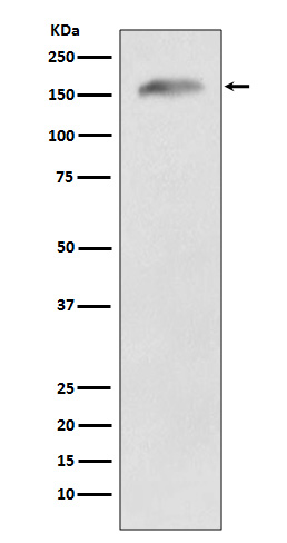
Western blot(SDS PAGE) analysis of extracts from 293T cell lysate.Using APC Rabbit mAb [T3p8]at dilution of 1:1000 incubated at 4℃ over night.
Product information
Protein names :APC; APC protein; Adenomatous polyposis coli protein; DP2.5; mAPC;
UniProtID :P25054
MASS(da) :311,646
MW(kDa) :160kDa
Form :Liquid
Purification :Affinity-chromatography
Host :Rabbit
Isotype : IgG
sensitivity :Endogenous
Reactivity :Human,Mouse,Rat
- ApplicationDilution
- 免疫印迹(WB)1:1000-2000
- 免疫组化(IHC)1:100
- 免疫荧光(ICC/IF)1:100
- The optimal dilutions should be determined by the end user
Specificity :Antibody is produced by immunizing animals with A synthesized peptide derived from human APC
Storage :Antibody store in 10 mM PBS, 0.5mg/ml BSA, 50% glycerol. Shipped at 4°C. Store at-20°C or -80°C. Products are valid for one natural year of receipt.Avoid repeated freeze / thaw cycles.
WB Positive detected :293T cell lysate.
Function : Tumor suppressor. Promotes rapid degradation of CTNNB1 and participates in Wnt signaling as a negative regulator. APC activity is correlated with its phosphorylation state. Activates the GEF activity of SPATA13 and ARHGEF4. Plays a role in hepatocyte growth factor (HGF)-induced cell migration. Required for MMP9 up-regulation via the JNK signaling pathway in colorectal tumor cells. Acts as a mediator of ERBB2-dependent stabilization of microtubules at the cell cortex. It is required for the localization of MACF1 to the cell membrane and this localization of MACF1 is critical for its function in microtubule stabilization..
Tissue specificity :Expressed in a variety of tissues: brain, small intestine, colon, thymus, skeletal muscle, heart, prostate, lung, spleen, ovary, testis kidney, placenta, blood and liver (PubMed:21643010, PubMed:27217144). Isoform 1A: Very strongly expressed in brain but has relatively low expression levels in other tissues (PubMed:19527921, PubMed:21643010, PubMed:27217144). Isoform 1B: Predominant form in all tissues except for brain, including gastric mucosa and blood (PubMed:19527921, PubMed:21643010, PubMed:27217144)..
Subcellular locationi :Cell junction, adherens junction. Cytoplasm, cytoskeleton. Cell projection, lamellipodium. Cell projection, ruffle membrane. Cytoplasm. Cell membrane.
IMPORTANT: For western blots, incubate membrane with diluted primary antibody in 1% w/v BSA, 1X TBST at 4°C overnight.


