
MUC1 Rabbit mAb [0KD0]Cat NO.: A44005
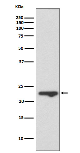
Western blot(SDS PAGE) analysis of extracts from T47 D cell lysate.Using MUC1 Rabbit mAb [0KD0]at dilution of 1:1000 incubated at 4℃ over night.
Product information
Protein names :MUC1; CA15-3; CD227; DF3 antigen; Episialin; Epithelial membrane antigen; H23 antigen; H23AG; Krebs von den Lungen-6; MAM6; MUC1/ZD; MUC-1/SEC; Mucin-1; Pem; KL-6; MUC-1/X; Tumor-associated mucin;
UniProtID :P15941
MASS(da) :122,102
MW(kDa) :18-25kDa
Form :Liquid
Purification :Affinity-chromatography
Host :Rabbit
Isotype : IgG
sensitivity :Endogenous
Reactivity :Human
- ApplicationDilution
- 免疫印迹(WB)1:1000-2000
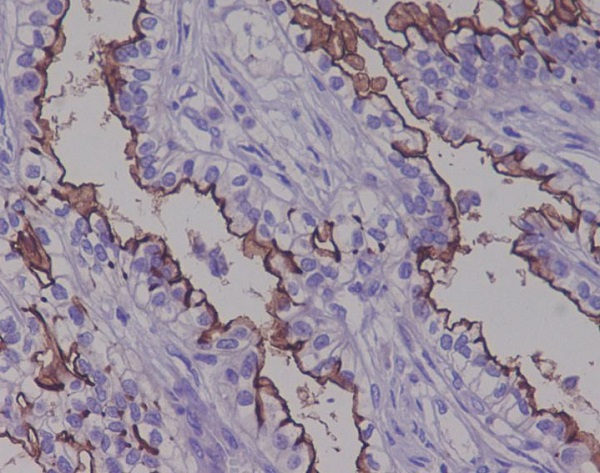
- 免疫组化(IHC)1:100
- 免疫荧光(ICC/IF)1:100
- The optimal dilutions should be determined by the end user
Specificity :Antibody is produced by immunizing animals with A synthesized peptide derived from human MUC1
Storage :Antibody store in 10 mM PBS, 0.5mg/ml BSA, 50% glycerol. Shipped at 4°C. Store at-20°C or -80°C. Products are valid for one natural year of receipt.Avoid repeated freeze / thaw cycles.
WB Positive detected :T47 D cell lysate.
Function : The alpha subunit has cell adhesive properties. Can act both as an adhesion and an anti-adhesion protein. May provide a protective layer on epithelial cells against bacterial and enzyme attack., The beta subunit contains a C-terminal domain which is involved in cell signaling, through phosphorylations and protein-protein interactions. Modulates signaling in ERK, SRC and NF-kappa-B pathways. In activated T-cells, influences directly or indirectly the Ras/MAPK pathway. Promotes tumor progression. Regulates TP53-mediated transcription and determines cell fate in the genotoxic stress response. Binds, together with KLF4, the PE21 promoter element of TP53 and represses TP53 activity.
Tissue specificity :Expressed on the apical surface of epithelial cells, especially of airway passages, breast and uterus. Also expressed in activated and unactivated T-cells. Overexpressed in epithelial tumors, such as breast or ovarian cancer and also in non-epithelial tumor cells. Isoform Y is expressed in tumor cells only..
Subcellular locationi :Apical cell membrane,Single-pass type I membrane protein.
IMPORTANT: For western blots, incubate membrane with diluted primary antibody in 1% w/v BSA, 1X TBST at 4°C overnight.
-
About
-
Product
-
Law
-
Literature
-

WeChat scan code follow us