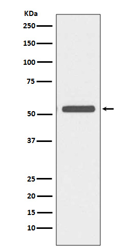
Cytokeratin 6 Rabbit mAb [U3UW]Cat NO.: A57492
Western blot(SDS PAGE) analysis of extracts from A431 cell lysate.Using Cytokeratin 6 Rabbit mAb [U3UW]at dilution of 1:1000 incubated at 4℃ over night.
Product information
Protein names :CK 6A; CK 6B; CK 6C; CK 6D; CK 6E; CK-6C; CK-6E; Cytokeratin 6a; Cytokeratin 6B; Cytokeratin 6C; Cytokeratin 6D; Cytokeratin 6E; Cytokeratin-6C; Cytokeratin-6E; K6a keratin; K6b keratin; K6C; K6c keratin; K6d keratin; K6e keratin; Keratin K6h;
UniProtID :P02538
MASS(da) :60,045
MW(kDa) :60kDa
Form :Liquid
Purification :Affinity-chromatography
Host :Rabbit
Isotype : IgG
sensitivity :Endogenous
Reactivity :Human
- ApplicationDilution
- 免疫印迹(WB)1:1000-2000
- 免疫组化(IHC)1:100
- 免疫荧光(ICC/IF)1:100
- The optimal dilutions should be determined by the end user
Specificity :Antibody is produced by immunizing animals with A synthesized peptide derived from human Cytokeratin 6
Storage :Antibody store in 10 mM PBS, 0.5mg/ml BSA, 50% glycerol. Shipped at 4°C. Store at-20°C or -80°C. Products are valid for one natural year of receipt.Avoid repeated freeze / thaw cycles.
WB Positive detected :A431 cell lysate.
Function : Epidermis-specific type I keratin involved in wound healing. Involved in the activation of follicular keratinocytes after wounding, while it does not play a major role in keratinocyte proliferation or migration. Participates in the regulation of epithelial migration by inhibiting the activity of SRC during wound repair..
Tissue specificity :Expressed in the corneal epithelium (at protein level)..
IMPORTANT: For western blots, incubate membrane with diluted primary antibody in 1% w/v BSA, 1X TBST at 4°C overnight.


